. 2024 Oct 30;13(21):1796.
doi: 10.3390/cells13211796.

Stress Granule Assembly in Pulmonary Arterial Hypertension

Affiliations

Stress Granule Assembly in Pulmonary Arterial Hypertension

Kosmas Kosmas et al. Cells. .

Abstract

The role of stress granules (SGs) in pulmonary arterial hypertension (PAH) is unknown. We hypothesized that SG formation contributes to abnormal vascular phenotypes, and cardiac and skeletal muscle dysfunction in PAH. Using the rat Sugen/hypoxia (SU/Hx) model of PAH, we demonstrate the formation of SG puncta and increased expression of SG proteins compared to control animals in lungs, right ventricles, and soleus muscles. Acetazolamide (ACTZ) treatment ameliorated the disease and reduced SG formation in all of these tissues. Primary pulmonary artery smooth muscle cells (PASMCs) from diseased animals had increased SG protein expression and SG number after acute oxidative stress and this was ameliorated by ACTZ. Pharmacologic inhibition of SG formation or genetic ablation of the SG assembly protein (G3BP1) altered the SU/Hx-PASMC phenotype by decreasing proliferation, increasing apoptosis and modulating synthetic and contractile marker expression. In human PAH lungs, we found increased SG puncta in pulmonary arteries compared to control lungs and in human PAH-PASMCs we found increased SGs after acute oxidative stress compared to healthy PASMCs. Genetic ablation of G3BP1 in human PAH-PASMCs resulted in a phenotypic switch to a less synthetic and more contractile phenotype. We conclude that increased SG formation in PASMCs and other tissues may contribute to PAH pathogenesis.

Keywords: ACTZ; Caprin1; G3BP1; ISRIB; PAH; stress granules; vascular smooth muscle cells.


Conflict of interest statement

L.E.F. is an employee of Regeneron Pharmaceuticals and holds stock and stock options. Other authors declare no conflicts of interest.

Figures

Figure 1
Figure 1
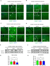
SG puncta in lungs from animals with SU/Hx-induced PH compared to controls and with ACTZ treatment. Paraffin-embedded sections of lungs stained with G3BP1. (A) G3BP1 puncta in pulmonary alveoli (upper panels) indicated by arrows; higher G3BP1 expression in airways (middle panels) and in pulmonary blood vessels (lower panels) in lungs from animals with SU/Hx-induced PH compared to controls and those with ACTZ treatment. Paraffin-embedded sections of lungs stained with Caprin1 (B) antibodies. Arrows indicate Caprin1 puncta, quantified with CellProfiler (C). Nuclei were visualized with DAPI staining. Scale bar, 20 μm. (D) Immunoblots showing upregulation of Caprin1 and no difference in G3BP1 expression levels in lung lysates of SU/Hx animals compared to SU/Hx-ACTZ-treated and controls, and quantitative analysis of immunoblots. (E) β-actin was used as a loading control, n = 3 animals per group; ns: not significant, * p < 0.05, *** p < 0.001, **** p < 0.0001.
Figure 2
Figure 2
Sarcoplasmic G3BP1 and Caprin1 puncta in right ventricles from animals with SU/Hx-induced PH compared to controls and with ACTZ treatment. Paraffin-embedded sections of right ventricles (longitudinal upper panels and transverse lower panels) (A); left ventricles (longitudinal upper panels and transverse lower panels) (B) stained with G3BP1 antibodies; right ventricles (longitudinal upper panels and transverse lower panels) (C); and left ventricles (longitudinal upper panels and transverse lower panels) (D) stained with Caprin1 antibodies. Arrows indicate G3BP1 and Caprin1 puncta respectively. Arrowheads indicate debris at the periphery of the cardiac muscle fibers. Nuclei were visualized with DAPI staining. Scale bar, 20 μm. (E) Immunoblots showing Caprin1 protein expression in right ventricles lysates of SU/Hx animals compared to SU/Hx-ACTZ treated and controls, and quantitative analysis of immunoblots (G). (F) Immunoblots showing Caprin1 protein expression in left ventricles lysates of SU/Hx animals compared to SU/Hx-ACTZ treated and controls, and quantitative analysis of immunoblots (H). Vinculin was used as a loading control, n = 6 animals per group; ns: not significant.
Figure 3
Figure 3
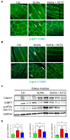
Sarcoplasmic G3BP1 and Caprin1 puncta in soleus muscles from animals with SU/Hx-induced PH compared to controls and with ACTZ treatment. Paraffin-embedded sections of soleus (longitudinal upper panels and transverse lower panels) stained with G3BP1 (A) and Caprin1 (B) antibodies. Arrows indicate G3BP1 and Caprin1 puncta, respectively. Nuclei were visualized with DAPI staining. Scale bar, 20 μm. (C) Immunoblots showing upregulation of SG components’ (Caprin1, G3BP1, p-eIF2α) protein expression in soleus lysates of SU/Hx animals compared to SU/Hx-ACTZ-treated animals and controls, and quantitative analysis of immunoblots (D). GAPDH was used as a loading control; n = 6 animals per group; ns: not significant.
Figure 4
Figure 4
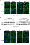
RPASMCs from SU/Hx animals have increased SG markers at baseline and more SGs after acute oxidative stress, and in vivo and in vitro treatment with ACTZ results in lower numbers of SGs. (A) Immunoblot showing upregulation of SG components (Caprin1 and G3BP1) in RPASMC lysates isolated from SU/Hx animals compared to controls. GAPDH was used as a loading control. (B) RPASMCs from SU/Hx animals, SU/Hx animals treated with ACTZ, and controls were treated with 0.5 mM arsenite for 40 min and stained for G3BP1 to detect SGs, quantified with CellProfiler. (C) (n = 55 cells/cell type). (D) RPASMCs were pre-treated with 1 mM ACTZ for 24 h, then treated with 0.5 mM arsenite for 40 min and stained for G3BP1 to detect SGs, quantified with CellProfiler (E) (n = 125 cells/cell type). Nuclei were visualized with DAPI staining. Scale bar, 20 μm; **** p < 0.0001. * p < 0.05.
Figure 5
Figure 5
ISRIB treatment results in lower numbers of SGs after oxidative stress and restores the contractile phenotype of RPASMCs from SU/Hx animals. (A) RPASMCs from SU/Hx animals and controls were pre-treated with ISRIB for 4 h, then treated with 0.5 mM arsenite for 40 min and stained for G3BP1 to detect SGs. Nuclei were visualized with DAPI staining. Scale bar, 20 μm. (B) Immunoblots showing downregulation of G3BP1 and slight decrease in p-eIf2α when pre-treated with ISRIB for 4 h, then treated with 0.5 mM arsenite for 40 min in RPASMCs isolated from SU/Hx animals compared to control cells. (C) RPASMCs from SU/Hx animals and controls were pre-treated with ISRIB for 4 h, then treated with 0.5 mM arsenite for 40 min and stained for αSMA to detect stress fibers and focal adhesions. Nuclei were visualized with DAPI staining. Scale bar, 20 μm.
Figure 6
Figure 6
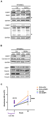
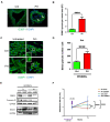
G3BP1 downregulation increases apoptosis, restores a contractile phenotype, and decreases proliferation of RPASMCs isolated from SU/Hx animals compared to control animals. (A) Immunoblots confirming downregulation of G3BP1 in RPASMCs isolated from SU/Hx animals and control cells. Knockdown of G3BP1 induces apoptosis (as assessed by cleaved PARP and cleaved caspase 3, indicated by stars) and, respectively, inhibits synthetic markers (Connexin 43) and increases contractile marker (desmin and α-SMA) expression in RPASMCs isolated from SU/Hx animals compared to control cells (B). (C) Knockdown of G3BP1 decreased the number of RPASMCs isolated from SU/Hx animals compared to control cells at 48 h (Cell proliferation reagent WST-1); **** p < 0.0001.
Figure 7
Figure 7
SG puncta in pulmonary blood vessels in lungs from patients with PAH compared to controls; PASMCs from the same patients have more SGs after acute oxidative stress and G3BP1 downregulation decreases their proliferation compared to controls. (A) Paraffin-embedded sections of lungs from patients with PAH and healthy donors stained with G3BP1. Arrows indicate G3BP1 puncta, quantified with CellProfiler (B). (C) PASMCs from patients with PAH and healthy donors were treated with 0.5 mM arsenite for 40 min and stained for G3BP1 to detect SGs, quantified with CellProfiler (D). Nuclei were visualized with DAPI staining. Scale bar, 20 μm; **** p < 0.0001 (n = 45 cells/cell type). (E) Immunoblots confirming downregulation of G3BP1 in hPASMCs isolated from patients with PAH and healthy donors. Knockdown of G3BP1 inhibits synthetic marker (Connexin 43) and increases contractile marker (desmin) expression in PASMCs isolated from patients with PAH and healthy donors. (F) Knockdown of G3BP1 decreased the number of HPASMCs isolated from patients with PAH compared to healthy donors at 72 h (Cell proliferation reagent WST-1); ** p < 0.01, **** p < 0.0001.

References

    1. Farber H.W., Loscalzo J. Pulmonary arterial hypertension. N. Engl. J. Med. 2004;351:1655–1665. doi: 10.1056/NEJMra035488. - DOI - PubMed
    1. Lechartier B., Berrebeh N., Huertas A., Humbert M., Guignabert C., Tu L. Phenotypic Diversity of Vascular Smooth Muscle Cells in Pulmonary Arterial Hypertension: Implications for Therapy. Chest. 2022;161:219–231. doi: 10.1016/j.chest.2021.08.040. - DOI - PubMed
    1. Hudalla H., Michael Z., Christodoulou N., Willis G.R., Fernandez-Gonzalez A., Filatava E.J., Dieffenbach P., Fredenburgh L.E., Stearman R.S., Geraci M.W., et al. Carbonic Anhydrase Inhibition Ameliorates Inflammation and Experimental Pulmonary Hypertension. Am. J. Respir. Cell Mol. Biol. 2019;61:512–524. doi: 10.1165/rcmb.2018-0232OC. - DOI - PMC - PubMed
    1. Kedersha N., Anderson P. Mammalian stress granules and processing bodies. Methods Enzymol. 2007;431:61–81. - PubMed
    1. Baymiller M., Moon S.L. Stress Granules as Causes and Consequences of Translation Suppression. Antioxid. Redox Signal. 2023;39:390–409. doi: 10.1089/ars.2022.0164. - DOI - PMC - PubMed

Publication types

MeSH terms

LinkOut - more resources