. 2024 Nov 8:13:RP101221.
doi: 10.7554/eLife.101221.

FABP4-mediated lipid accumulation and lipolysis in tumor-associated macrophages promote breast cancer metastasis

Affiliations

FABP4-mediated lipid accumulation and lipolysis in tumor-associated macrophages promote breast cancer metastasis

Matthew Yorek et al. Elife. .

Abstract

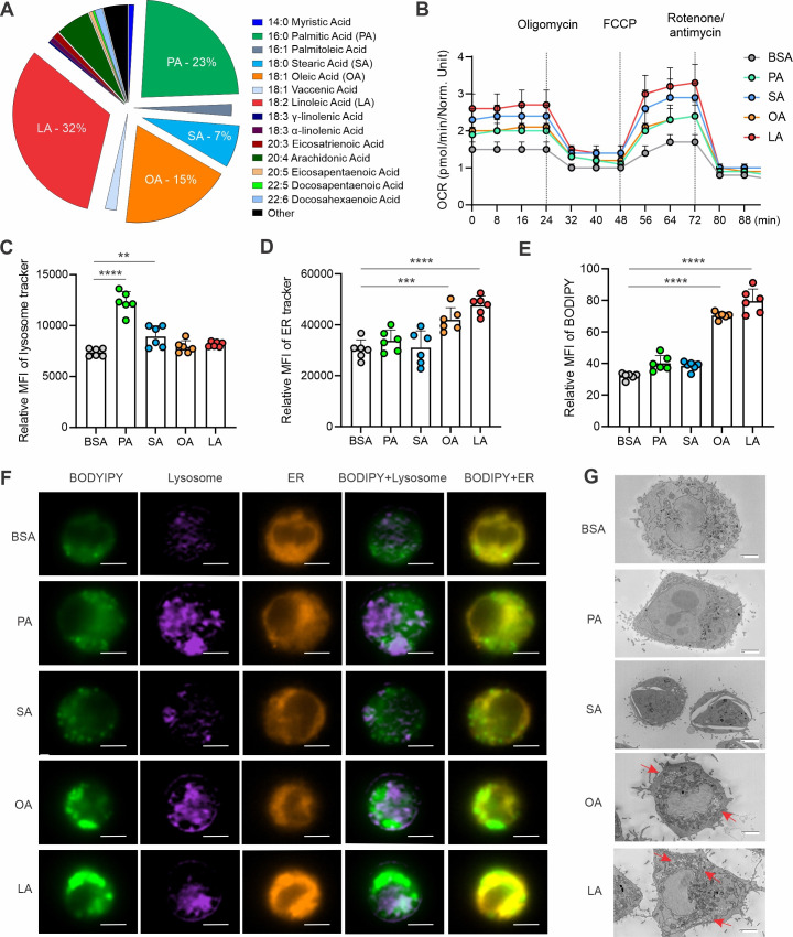
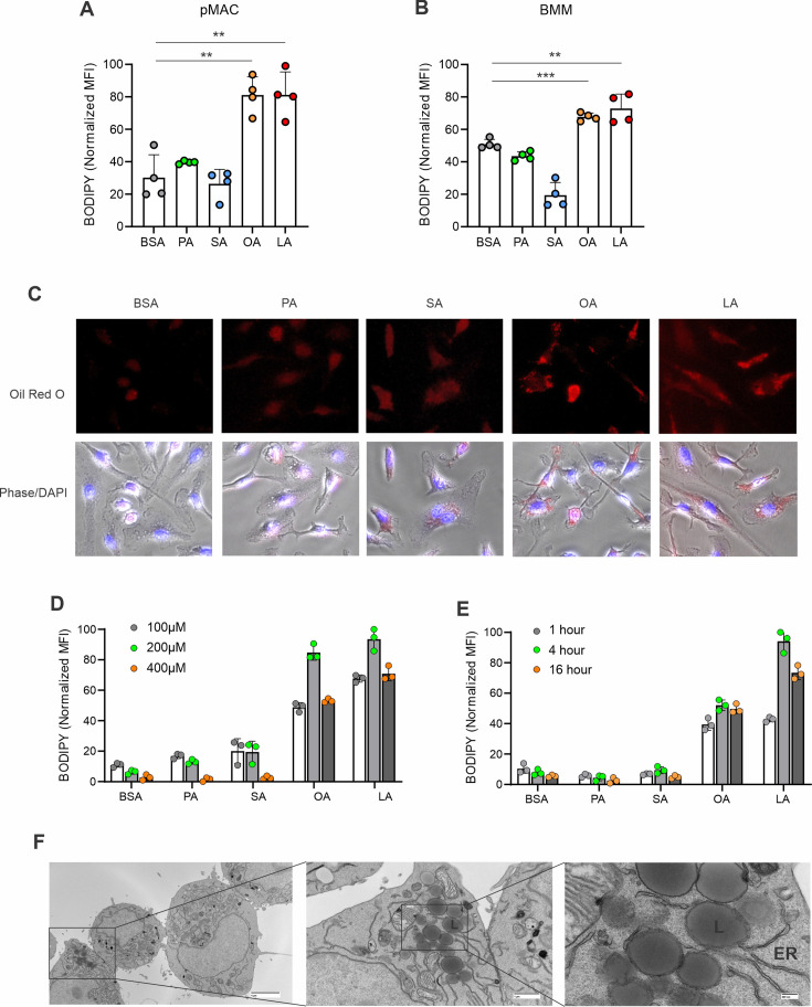
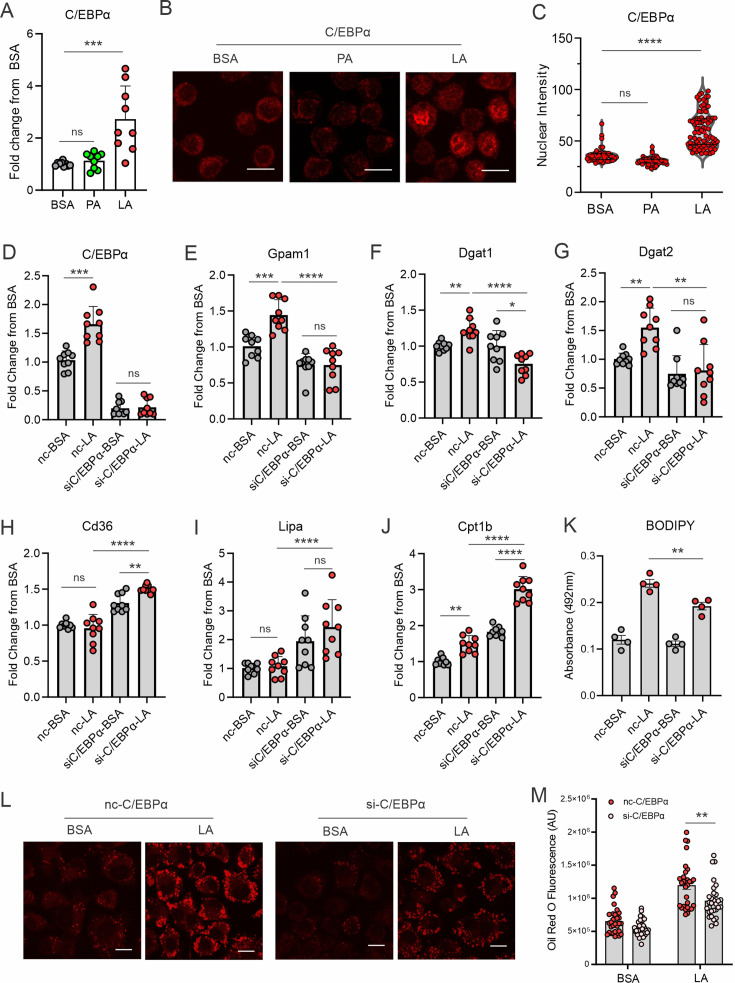
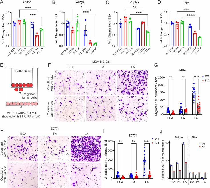
A high density of tumor-associated macrophages (TAMs) is associated with poorer prognosis and survival in breast cancer patients. Recent studies have shown that lipid accumulation in TAMs can promote tumor growth and metastasis in various models. However, the specific molecular mechanisms that drive lipid accumulation and tumor progression in TAMs remain largely unknown. Herein, we demonstrated that unsaturated fatty acids (FAs), unlike saturated ones, are more likely to form lipid droplets in murine macrophages. Specifically, unsaturated FAs, including linoleic acids (LA), activate the FABP4/CEBPα pathway, leading to triglyceride synthesis and lipid droplet formation. Furthermore, FABP4 enhances lipolysis and FA utilization by breast cancer cell lines, which promotes cancer cell migration in vitro and metastasis in vivo. Notably, a deficiency of FABP4 in murine macrophages significantly reduces LA-induced lipid metabolism. Therefore, our findings suggest FABP4 as a crucial lipid messenger that facilitates unsaturated FA-mediated lipid accumulation and lipolysis in TAMs, thus contributing to the metastasis of breast cancer.

Keywords: FABP4; breast cancer; cancer biology; human; lipid droplet formation; metastasis; mouse; tumor associated macrophages; unsaturated fats.


Conflict of interest statement

MY, XJ, SL, JH, JY, AA, ZL, MC, HK, JS, AK, MK, YX, ES, SS, BL No competing interests declared

Figures

Figure 1.
Figure 1.. Unsaturated FAs form lipid accumulation in macrophages.
(A) Pie chart showing main compositions of free fatty acids in the serum of healthy humans. (B) Measurement of oxygen consumption rate (OCR) in macrophages treated with 200 μM of PA, SA, OA, LA or control BSA, respectively, under basal conditions or following the addition of oligomycin, FCCP or the electron transport inhibitor Rotenone/antimycin by a seahorse XF-96 analyzer (n=5). (C–E) Macrophages were treated with 200 μM of PA, SA, OA, LA or BSA for 4 hr. Flow cytometric analysis of lysosome, ER and lipid droplet formation by measuring mean fluorescent intensity (MFI) of lysosome tracker (C), ER tracker (D) and BODIPY (E) in macrophages. (F) Multispectral imaging analysis of BODIPY (green), Lysosome (purple), ER (orange), and merged images showing the colocalization status of BODIPY/lyso-tracker and BODIPY/ER-tracker in macrophages treated with PA, SA, OA, LA, or BSA at 200 μM for 4 hr. Scale bar: 7μM. (G) Analysis of lipid droplet formation (red arrow) in macrophages treated with BSA, PA, SA, OA, and LA by transmission electron microscope. Data are shown as mean ± SD in panel B-E (** p≤0.01, *** p≤0.001, **** p≤0.0001, as compared to the control BSA group, unpaired Student t test).
Figure 1—figure supplement 1.
Figure 1—figure supplement 1.. Unsaturated FA form lipid droplets in different macrophages.
(A) Flow cytometric analysis of lipid droplet formation in peritoneal macrophages treated with BSA and indicated dietary FA (100 μM). (B) Flow cytometric analysis of lipid droplet formation in bone-marrow-derived macrophages (BMM) treated with BSA and indicated dietary FA (100 μM). (C) Confocal microscopy analysis of Oil Red O staining in BMM treated with 200 μM of PA, SA, OA, LA, and control BSA for 4 hr. (D) Titration of different indicated concentrations of dietary FA in forming lipid droplets in macrophage cell lines. (E) Measurement of lipid droplet formation in macrophages treated with indicated time periods of different dietary FAs (100 μM). (F) Transmission electron microscope (TEM) images showing that LA-induced lipid droplets were formed in endoplasmic reticulum (ER) in macrophages. Data are shown as mean ± SD in panel (** p≤0.01, *** p≤0.001, unpaired Student t test).
Figure 2.
Figure 2.. LA induces the expression of key enzymes of triacylglycerol synthesis in macrophages.
(A) Key enzymes in the biosynthesis of triacylglycerol. (B–H) Analyzing the expression of genes encoding key enzymes, including Gpam1 (B), Gpam3 (C), Gpam4 (D), Agpat2 (E), Lipin1a (F), Dgat1 (G), Dgat2 (H), in the triacylglycerol biosynthesis pathway in macrophages treated with 400 μM of PA, LA or BSA for 4 hr by real-time PCR. (I) Representative confocal images of lipid accumulation by LipidTOX staining (green), expression of GPAT1 (red), DGAT1 in macrophages treated with BSA, PA, and LA (400 μM) overnight. (J–M) Quantification of lipid accumulation (J), protein levels of GAPT1 (K) and DGAT1 (M) in macrophages treated with BSA, PA, or LA (400 μM) overnight. Data are shown as mean ± SD in panel B-H, J-M (** p≤0.01, *** p≤0.001, **** p≤0.0001, ns, non-significant, as compared to the control BSA group, unpaired Student t test).
Figure 2—figure supplement 1.
Figure 2—figure supplement 1.. LA induces key enzyme expression in triglyceride biosynthesis.
(A–F) Analysis of FA metabolism-related genes, including Acs1 (A), Cpt1a (B), Cpt1b (C), HMGCR (D), Acat1 (E), Acat2 (F) in macrophages treated with BSA, PA or LA (400 μM) for 4 hr. (G–I) Flow cytometric staining for LipidTOX (G), GPAT1 (H), and DGAT1 (I) expression in macrophages treated with BSA, PA, or LA (400 μM). (J–L) Quantification of LipiTOX (J), GPAT1 (K), and DGAT1 (L) expression by mean fluorescent intensity (MFI) in macrophages treated with BSA, PA, or LA (400 μM). Data are shown as mean ± SD in panel A-F and J-L (* p≤0.05, ** p≤0.01, **** p≤0.0001, ns, non-significant, unpaired Student t test).
Figure 3.
Figure 3.. LA induces lipid accumulation through activating the C/EBPα pathway.
(A) Measurement of C/EBPα gene expression levels in macrophages treated with BSA, PA, or LA (400 μM) for 4 hr by real-time PCR. (B) Representative confocal images of C/EBPα protein expression (red) in macrophages treated with BSA, PA, or LA overnight. (C) Quantification of C/EBPα nuclear expression in macrophages treated with BSA, PA, or LA overnight by Image J analysis. (D–J) Real-time PCR analysis of the levels of C/EBPα (D), Gpam1 (E), Dgat1 (F), Dgat2 (G), Cd36 (H), Lipa (I), and Cpt1b (J) in macrophages transfected with 40 nM C/EBPα siRNA or control siNC and then treated with BSA or LA for 4 hr. (K) Measurement of BODIPY fluorescent intensity in C/EBPα-silencing or control macrophages treated with BSA or LA using a fluorescence spectroscopy. (L) Representative confocal images of Oil Red O staining in C/EBPα-silencing or control macrophages treated with BSA or LA (bar, 10 μM). (M) Quantification of Oil Red O fluorescence intensity in C/EBPα-silencing or control macrophages treated with BSA or LA. Data are shown as mean ± SD in panel A, C-K and M (*p≤0.05, ** p≤0.01, *** p≤0.001, **** p≤0.0001, ns, non-significant, as compared to the control BSA group or control siNC group, unpaired Student t test).
Figure 3—figure supplement 1.
Figure 3—figure supplement 1.. LA-induced lipid accumulation in macrophages was undependable on CEBPβ.
(A) CiiiDER software package was used to identify potential C/EBPα and C/EBPβ transcription factor binding sites in regulatory regions of relevant lipid droplet and lipolysis genes. (B) Real-time PCR analysis of CEBPβ expression macrophages treated with BSA, PA, and LA for 4 hr. (C, D) Confocal analysis for CEBPβ staining (green) in macrophages treated with BSA, PA, and LA. Quantification of CEBPβ staining is shown in panel D. (E–K) Macrophages was transfected with CEBPβ or control siRNA, and then treated with BSA or LA for 4 hr. Expression of CEBPβ (E), lipid droplet formation and other FA-metabolism related genes, including Gpam1 (F), Cd36 (G), Lipa (H), Dgat1 (I), Dgat2 (J), Cpt1b (K) was determined by real-time PCR analysis. (L) Measurement of lipid droplet formation by BODIPY staining in CEBPβ-silencing macrophages treated with BSA or LA. Data are shown as mean ± SD in panel L-O (** p≤0.01, **** p≤0.0001, ns, non-significant, unpaired Student t test).
Figure 4.
Figure 4.. FABP4 mediates LA-induced C/EBPα expression in macrophages.
(A–C) Analysis of the expression of FABP family members, including Fabp3 (A), Fabp4 (B) and Fabp5 (C), in macrophages treated with BSA, PA or LA (400 μM) for 4 hours. (D) UMAP of FABP4-positive macrophage subsets using mouse spleen single-cell RNA sequence analysis. (E–J) Violin plots showing relative expression levels of genes, including Fabp4(E), Cd36 (F), PPARγ (G), CEBPA (H), Plin2 (I) and Fabp5 (J) between Fabp4 +vs Fabp4- macrophages indicated in (C). (K) Confocal analysis of FABP4 expression in macrophages treated with BSA, PA or LA (400 μM) (bar, 10 μM). (L) Comparison of the expression of FABP4 and C/EBPα between FABP4 wildtype (WT) and knockout (KO) macrophages in response to LA treatment (400 μM) (bar, 10 μM). (M) Realtime PCR analysis of CEBPA expression in WT and FABP4-/- macrophages treated with BSA, PA, and LA (400 μM). Data are shown as mean ± SD in panel A and L (** p≤0.01, *** p≤0.001, as compared to the control BSA group or FABP4-/- group, unpaired Student t test).
Figure 4—figure supplement 1.
Figure 4—figure supplement 1.. FABP4 mediates LA-induced CEBPα, but not CEBPβ, activation.
(A, B) Spearman’s correlation analysis of CEBPα with FABP family members, especially FABP4 with CEBPα (B), using cBioportal breast cancer TCGA database. (C) Spearman’s correlation analysis of FABP4 with CEBP family members using cBioportal breast cancer TCGA database. (D–F) Differential gene expression analysis of FABP4, GPAM, DGAT1, DGAT2, CEBPA, and CD36 in published breast cancer microarray databases, including GSE42568 (D), GSE21422 (E) and GSE29431 (F). (G) UMAP of different subsets of splenic macrophages using single-cell RNA sequence analysis. (H) Molecular function pathway analysis of the integrated conserved genes in the FABP4-positive clusters by g:Profiler. (I–L) Violin plots showing relative expression levels of genes, including Acs1(I), Lipa (J) and Fasn (K) and Cpt1b (L) between FABP4 +vs FABP4- macrophages in the splenic macrophage single cell RNA sequencing analysis. (M) Measurement of CEBPA gene levels in WT and FABP4-/- peritoneal macrophages treated with BSA, PA, or LA, respectively, for 4 hr. (N) Measurement of CEBPA gene levels in WT and FABP4-/- bone-marrow-derived macrophages treated with BSA, PA, or LA, respectively, for 4 hr. (O) Measurement of CEBPB gene levels in WT and FABP4-/- peritoneal macrophages treated with BSA, PA, or LA, respectively, for 4 hr. (P) Measurement of CEBPB gene levels in WT and FABP4-/- bone-marrow-derived macrophages treated with BSA, PA, or LA, respectively, for 4 hr. Data are shown as mean ± SD in panels I-P (p* p≤0.05, ** p≤0.01, ns, non-significant, unpaired Student t test).
Figure 5.
Figure 5.. FABP4 deficiency reduces LA-induced lipid accumulation in macrophages.
(A–F) real-time PCR analysis of FABP4 (A) and genes encoding key enzymes for triglycerol biosynthesis, including Gpam1 (B), Dgat1 (C), Dgat2 (D), Gpam4 (E), Agpat2 (F) in WT and FABP4 KO macrophages treated with BSA, PA, or LA (400 μM). (G) Confocal analysis of protein expression of GPAT1 (red), DGAT1 (cyan) and lipid accumulation (LipidTOX staining, green) in LA-treated WT and FABP4 KO macrophages (bar, 10 μM). (H–J) Expression levels of GPAT1 (H), DGAT1 (I) and LipidTOX (J) as indicated in panel G were quantified by Image J. (L) Flow cytometric analysis of neutral lipid accumulation as shown by BODIPY staining in WT and FABP4 KO macrophages treated with BSA or LA. (M) Transmission electron microscope showing lipid droplet staining in WT and FABP4 KO macrophages treated with LA. Data are shown as mean ± SD in panels A-F, H-L (* p≤0.01, p** p≤0.01, *** p≤0.001, ns, non-significant as compared to the control BSA group or FABP4-/- group, unpaired Student t test).
Figure 5—figure supplement 1.
Figure 5—figure supplement 1.. Deficiency of FABP4 reduces lipid droplet formation in macrophages.
(A–F) Real-time PCR analysis of FABP4 (A) and genes encoding key enzymes for triglycerol biosynthesis, including Gpam1 (B), Dgat1 (C), Dgat2 (D), Cd36 (E), Fabp5 (F) in WT and FABP4 KO peritoneal macrophages treated with BSA, PA, and LA (400 μM). (G) Transmission electron microscope showing lipid droplet formation (red arrows) in WT and KO macrophages treated with either PA or OA for 4 hr. (H, I) Flow cytometric analysis of BODIPY staining (G) and mean fluorescent intensity (MFI) in peritoneal macrophages treated with BSA, PA, or LA (400 μM). Data are shown as mean ± SD (*** p≤0.001, *** p≤0.001, unpaired Student t test).
Figure 6.
Figure 6.. FAPB4 expression in macrophages promotes lipolysis and breast cancer cell migration.
(A–D) Realtime PCR analysis of expression of Adrb2 (A), Adcy4 (B), Pnpla2 (C) and Lipe (D) in FABP4 WT and KO macrophages treated with BSA, PA, or LA (400 μM). (E) Transwell measurement of migration of breast cancer cells cocultured with FA- or BSA-treated FABP4 WT or KO macrophages (Mφ). (F–I) FABP4 WT or KO macrophages were treated with 100 μM BSA, PA, or LA for 4 hr. Fatty acids in the culture medium were washed away with FBS-free RPMI-1640. Breast cancer cells were added to a transwell and cocultured with these different FA-or BSA-treated FABP4 WT or KO macrophages for 24 hr. The migrated tumor cells were stained and quantified. Migrated MDA-MB-231 cells were shown in panels F and G. Migrated E0771 cells were shown in panels H and I. (J) FABP4 WT and KO macrophages were treated with indicated FAs or BSA for 4 hr. Flow cytometric staining of BODIPY levels in WT and KO macrophages before and after coculture with E0771 tumor cells for 24 hr. Data are shown as mean ± SD in panels A-D, G, I, J (* p≤0.01, p** p≤0.01, *** p≤0.001, ns, non-significant as compared to the control group or FABP4 KO group, unpaired Student t test).
Figure 6—figure supplement 1.
Figure 6—figure supplement 1.. FABP4 promotes lipolysis and tumor migration.
(A–D) Spearman’s correlation analysis between FABP4 and key genes mediating lipolysis, including Adrb2 (A), Adcy4 (B), Pnpla2 (C), and Lipe (D) using the cBioportal breast cancer TCGA database. (E) Measurement of cell proliferation marker Ki67 in E0771 tumor cells after coculture with FA-treated FABP4 WT or KO macrophages for 24 hr. (F, G) After treatment with BSA, PA, or LA (400 μM) for 4 hr, FABP4 WT and KO macrophages were cultured in FBS-free RPMI-1640 for 24 hr. The cultural conditional medium was collected for tumor migration assays. MDA-MB-231 migration and quantification are shown in panels F and G, respectively. (H, I) E0771 tumor migration assays were performed using the conditional medium collected as above plus 1% FBS. Tumor migration and quantification are shown in panels H and I, respectively. (J) ELSIA measurement of FABP4 levels in macrophage/tumor coculture medium. Data are shown as mean ± SD in panels G, I, and J (p* p≤0.05, *** p≤0.001, ns, non-significant, unpaired Student t test).
Figure 7.
Figure 7.. High expression of FABP4 in TAMs is associated with more metastasis of breast cancer.
(A) Comparison of H&E and CD163 staining (brown) between an example of small and large breast cancer tumors in breast cancer patients. (B) Spearman correlation analysis between breast cancer tumor size and CD163 +TAM staining. (C) Expression of FABP4 and CD163 was highly correlated as analyzed by the Spearman correlation analysis in breast cancer tissues. (D) Pie chart showing the percentage of breast cancer patients with or without metastasis. (E) Analysis of the staining of H&E, CD163 (brown), FABP4 (red) in primary breast tumors of patients with and without metastasis. (F) Analysis of CD163 expression levels between primary breast tumors of patients with and without metastases. (G) Analysis of FABP4 expression levels between primary breast tumors of patients with and without metastases. (H) Analysis of FABP4 expression levels between alive and deceased breast cancer patients. (I) Scheme of how FABP4 mediates unsaturated FA (yellow)-induced lipid storage and lipolysis in TAMs. When TAMs are exposed to dietary saturated (gray) or unsaturated (yellow) FAs, unsaturated FAs, but not saturated ones, induce FABP4 nuclear translocation and upregulate FABP4 and CEBPA-mediated transactivation of GPAM1 and DGATs, promoting lipid storage as lipid droplets. Once tumor-induced lipolysis occurs, FABP4/unsaturated FAs are secreted from TAMs to induce tumor migration and metastasis. Data are shown as mean ± SD in panel F-H (p** p0.01, *** p≤0.001, *** p≤0.001, unpaired Student t test).
Figure 7—figure supplement 1.
Figure 7—figure supplement 1.. Association of TAMs with obesity and survival of breast cancer patients.
(A–D) PROGgene analysis of the association between CD163 expression levels and breast cancer survival using various publicly accessible breast cancer databases, including NKI (A), GSE10893 (B), GSE18229 (C) and GSE9893 (D). (E) Comparison of H&E and CD163 staining (brown) between lean and obese breast cancer tumors in breast cancer patients. (F) Spearman’s correlation analysis between CD163 expression in breast cancer tissues and BMI at diagnosis of breast cancer patients. (G) H&E and immunohistochemistry staining of FABP4 (red) in ER +and triple negative breast cancer tissues. (H) Analysis of FABP4 expression in hormone receptor+/HER2-, HER2 +and triple negative breast cancers. Data are shown as mean ± SD in panel H (p* p≤0.05, ** p≤0.01, ns, non-significant, unpaired Student t test).
Author response image 1.
Author response image 1.. LA induced PPARg expression in macrophages.
Bone-marrow derived macrophages were treated with 400μM saturated FA (SFA), unsaturated FA (UFA) or BSA control for 6 hours. PPARg expression was measured by qPCR (***p<0.001).

Update of

References

    1. Allison E, Edirimanne S, Matthews J, Fuller SJ. Breast cancer survival outcomes and tumor-associated macrophage markers: A systematic review and meta-analysis. Oncology and Therapy. 2023;11:27–48. doi: 10.1007/s40487-022-00214-3. - DOI - PMC - PubMed
    1. Arnold M, Morgan E, Rumgay H, Mafra A, Singh D, Laversanne M, Vignat J, Gralow JR, Cardoso F, Siesling S, Soerjomataram I. Current and future burden of breast cancer: Global statistics for 2020 and 2040. Breast. 2022;66:15–23. doi: 10.1016/j.breast.2022.08.010. - DOI - PMC - PubMed
    1. Batista-Gonzalez A, Vidal R, Criollo A, Carreño LJ. New insights on the role of lipid metabolism in the metabolic reprogramming of macrophages. Frontiers in Immunology. 2019;10:2993. doi: 10.3389/fimmu.2019.02993. - DOI - PMC - PubMed
    1. Beck AH, Espinosa I, Edris B, Li R, Montgomery K, Zhu S, Varma S, Marinelli RJ, van de Rijn M, West RB. The macrophage colony-stimulating factor 1 response signature in breast carcinoma. Clinical Cancer Research. 2009;15:778–787. doi: 10.1158/1078-0432.CCR-08-1283. - DOI - PMC - PubMed
    1. Chen D, Wirth KM, Kizy S, Muretta JM, Markowski TW, Yong P, Sheka A, Abdelwahab H, Hertzel AV, Ikramuddin S, Yamamoto M, Bernlohr DA. Desmoglein 2 functions as a receptor for fatty acid binding protein 4 in breast cancer epithelial cells. Molecular Cancer Research. 2023;21:836–848. doi: 10.1158/1541-7786.MCR-22-0763. - DOI - PMC - PubMed