. 2024 Nov 12;121(46):e2408939121.
doi: 10.1073/pnas.2408939121. Epub 2024 Nov 8.

Lamination-based organoid spatially resolved transcriptomics technique for primary lung and liver organoid characterization

Affiliations

Lamination-based organoid spatially resolved transcriptomics technique for primary lung and liver organoid characterization

Shaohua Ma et al. Proc Natl Acad Sci U S A. .

Abstract

Spatial-transcriptomics technologies have demonstrated exceptional performance in characterizing brain and visceral organ tissues, as well as brain and retinal organoids. However, it has not yet been proven whether spatial transcriptomics can effectively characterize primary tissue-derived organoids, as the standardized tissue sectioning or slicing methods are not applicable for such organoids. Herein, we present a technique, lamination-based organoid spatially resolved transcriptomics (LOSRT), for organoid-spatially resolved transcriptomics based on organoid lamination. Primary mouse lung and liver-derived organoids were used in this study. The organoids were formulated using the droplet-engineering method and laminated using a homemade device with weight compression. This technique preserved most cells in individual organoids while maintaining delicate epithelium structures in laminated domains that can be recognized through visual segmentation. The mouse lung and liver organoids were resolved comprising various cell types, including alveolar cells, damage-associated transient progenitor cells, basal cells, macrophages, endothelial cells, fibroblasts, hepatocytes, and hepatic stellate cells. The distribution and count of cells were confirmed using immunohistology and identified with spatial transcriptomic features. This study reports an automated and integrated spatial transcriptomics method for primary organoids. It has the potential to standardize and rapidly characterize primary tissue-derived organoids.

Keywords: organoid; organoid lamination; spatial transcriptomics.


Conflict of interest statement

Competing interests statement:The authors declare no competing interest.

Figures
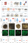
Fig. 1.
Fig. 1.
LOSRT technique. (A) Schematic of LOSRT. DEO, droplet-engineered organoid. DEO organoids are laminated in a device by weight compression and transferred to BGI Stereo-seq chips for spatial transcriptomics analysis. (B) Stepwise description of organoid lamination. (C) Preservation of the epithelial structure of mouse lung and liver organoids before (brightfield) and after (calcium AM-labeled fluorescence) organoid lamination. (D) Statistics of laminated mouse lung and liver DEO organoids, by quantifying the laminated area, cell number, cell symmetry, and cell dispersion. (E) Comparison of laminated organoids by using manual compression and device compression.
Fig. 2.
Fig. 2.
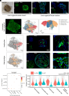
Spatially resolved transcriptome of mouse lung organoids using LOSRT. (A) Bright-field and live (Calcium AM)/dead (PI) cell whole mount imaging of a lung organoid and nuclei (DAPI), macrophages (F4/80), and goblet cells (Mu5ac) imaging of cryosectioned lung organoid sections. (B) ssDNA imaging, spatially resolved cell type annotation, and cell type classification of a laminated lung organoid. Orange: alveolar pneumocyte type I. Brown: alveolar pneumocyte type II. Green: basal cells. Blue: damage-associated transient progenitor cells. Purple: endothelial cells. Red: fibroblasts. Yellow: macrophages. (C) Spatially resolved distribution of alveolar type II cells labeled by the SFTPC gene and verified by immunofluorescence staining of SFTPC gene in cryosectioned organoid sections. (D) Spatially resolved distribution of macrophages labeled by F4/80 and verified by immunofluorescence staining of F4/80 in cryosectioned organoid sections. (E) Dot plot showing the spatial pattern of the laminated lung organoid. (F) Violin plot showing the spatial distance from different cell types in laminated lung organoids. Wilcoxon rank test was applied. *above the hepatocytes chart represents the significant difference between “to_other” and “to_self” groups with the P-value < 0.05.
Fig. 3.
Fig. 3.
Spatially resolved transcriptome of mouse liver organoids using LOSRT. (A) Bright-field and live (Calcium AM)/dead (PI) cell whole mount imaging of a liver organoid and nuclei (DAPI), hepatocytes (Hnf4α), and epithelial cells/cholangiocytes (Epcam) imaging of cryosectioned liver organoid sections. (B) ssDNA imaging, spatially resolved cell type annotation and cell type classification of a laminated liver organoid. Blue: endothelial cells. Orange: epithelial cells. Green: hepatic stellate cells. Red: hepatocytes. Yellow: macrophages. (C) Spatially resolved distribution of hepatocytes labeled by CK18 and verified by immunofluorescence staining of CK18 in cryosectioned organoid sections. (D) Spatially resolved distribution of macrophages labeled by CD68 and verified by immunofluorescence staining of CD68 in cryosectioned organoid sections. (E) Dot plot showing the spatial pattern of the laminated liver organoid. (F) Violin plot showing the spatial distance from different cell types in laminated liver organoids. Wilcoxon rank test was applied. *above the hepatocytes chart represents the significant difference between “to_other” and “to_self” groups with the P-value < 0.05.
Fig. 4.
Fig. 4.
Reproducible application of primary organoids in spatial transcriptome. (A) Three replicates of lamination of primary mouse lung organoids on one single transcriptomic profiling chip. The Right figure shows distribution of different cell types in each group of organoids resolved from the three organoids. (B) Another spatial transcriptomic result of primary mouse lung organoids. The Right figure shows distribution of different types of cells in the organoids (compared with cell distribution of Fig. 4A).

References

    1. Zhao Z., et al. , Organoids. Nat. Rev. Methods Primers 2, 94 (2022). - PMC - PubMed
    1. Polak R., Zhang E. T., Kuo C. J., Cancer organoids 2.0: Modelling the complexity of the tumour immune microenvironment. Nat. Rev. Cancer 24, 523–539 (2024). - PubMed
    1. Jiang S., et al. , An automated organoid platform with inter-organoid homogeneity and inter-patient heterogeneity. Cell Rep. Med. 1, 100161 (2020). - PMC - PubMed
    1. Chrisnandy A., Blondel D., Rezakhani S., Broguiere N., Lutolf M. P., Synthetic dynamic hydrogels promote degradation-independent in vitro organogenesis. Nat. Mater. 21, 479–487 (2022). - PubMed
    1. Chen A., et al. , Spatiotemporal transcriptomic atlas of mouse organogenesis using DNA nanoball-patterned arrays. Cell 185, 1777–1792.e21 (2022). - PubMed

LinkOut - more resources