. 2024 Dec 20;5(4):103439.
doi: 10.1016/j.xpro.2024.103439. Epub 2024 Nov 6.

Protocol for isolating specific C. elegans neuron types for bulk and single-cell RNA sequencing

Affiliations

Protocol for isolating specific C. elegans neuron types for bulk and single-cell RNA sequencing

Seth R Taylor et al. STAR Protoc. .

Abstract

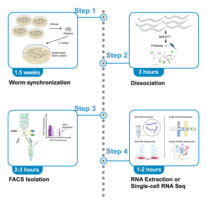
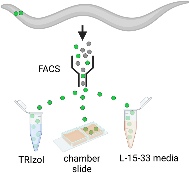
The C. elegans nervous system is compact and well described, ideal for identifying genetic programs that drive neuron-specific development, function, and connectivity. Here, we present a protocol for isolating specific neuron types for gene expression profiling by bulk or single-cell RNA sequencing. We describe steps for worm synchronization, dissociation, and fluorescence-activated cell sorting (FACS) isolation. We then detail procedures for RNA extraction and preparing cells for single-cell sequencing. This protocol is applicable to the isolation of individual cell types from larval and adult animals. For additional details on the use and execution of this protocol, see Taylor et al.1.

Keywords: cell biology; genomics; model organisms; neuroscience.


Conflict of interest statement

Declaration of interests The authors declare no competing interests.

Figures

None
Graphical abstract
Figure 1
Figure 1
Generating large volumes of synchronized worms (A) Expanding C. elegans cultures to yield hundreds of thousands to millions of synchronized worms. (B) Top: Worm culture plates with diameters of 60 mm (left), 100 mm (middle) and 150 mm (right) used in expanding the cultures. Bottom: Low-magnification view of a 150 mm plate ready for bleaching. Note (inset) the “windrow” of gravid adults (arrow). (C) Stereoscopic image of abundant gravid adults ready for bleaching. Scale bar is 200 microns.
Figure 2
Figure 2
SDS-DTT treatment weakens the cuticle for subsequent pronase digestion (A) Young adult worms after treatment with SDS-DTT for 2 min (left) or 4 min (right). (B) Bright-field images of young adult worms after incubation with pronase and 2 rounds of pipetting (Left: 2 min SDS-DTT, Right: 4 min SDS-DTT). (C) Dissociated GFP+ cells (arrowheads) after 10 min of pronase and 2 rounds of pipetting following 2-min SDS-DTT (left) or following 4-min SDS-DTT (right) treatment. Scale bars are 100 microns.
Figure 3
Figure 3
Using FACS to isolate fluorescently labeled neurons (A) Schematic of left-hand member (NSML) of NSML/R pharyngeal neuron pair, (WormAtlas). (B) Confocal image of tph-1::GFP expression in NSM and dimly labeled ADF neuron. (C) FACS isolation of GFP-labeled NSM neurons. (D and E) FACS profiles of cells dissociated from (D) tph-1::GFP and (E) N2 (control) strains depicting Side Scatter (SSC) vs. GFP fluorescence. (F–H) FACS light scattering plots. SSC & FSC gates (magenta) encompass tph-1::GFP-labeled cells (dark purple dots). (I) Bright DAPI fluorescence labels a population of damaged/dying cells vs. viable tph-1::GFP marked cells (box). (J) DAPI vs. GFP profile of control (N2) cells. Arrowheads denote target tph-1::GFP cells in FACS plots. (K) Table showing the fraction of events at each stage of gating, including percent of the previous gate (%Parent) and percent of all events (%Total), GFP+ = Panel 3D (arrowhead); GFP_+ = Panel 3I (arrowhead) is defined by a more stringent gate derived from the FSC pulse geometry profile (3H) and thus contains fewer cells than the GFP+ population (3D) identified at an earlier step of setting FACS parameters for the sort. (L) NSM neurons marked with tph-1::GFP cultured for 2–3 h after dissociating from young adult animals. Scale bars are 5 microns.
Figure 4
Figure 4
Sorting GFP-labeled neurons for RNA isolation (TRIzol), cell culture (chamber slide) or scRNA-Seq (L-15-33)
Figure 5
Figure 5
RNA Bioanalysis profiles from individual neuron classes (A) Bioanalysis (Agilent) profiles of high quality RNA samples extracted from sorts of individual neuron classes. Note RNA Integrity Numbers (RINs) from 8.1–9.3 and RNA yields of 200–300 pg/μL. (B) Bioanalysis profiles from lower quality RNA samples extracted from sorts of individual neuron classes. RINs from 1.2–7.5, RNA yields from 30–333 pg/μL.
Figure 6
Figure 6
Counting cells with hemocytometer
Figure 7
Figure 7
FACS yield and RNA yield show weak correlation Data from 219 RNA preparations extracted from sorts of individual neuron classes. 197 RNA preparations were obtained from single FACS isolations (“single”, blue) and 22 RNA preparations were extracted from samples pooled from multiple sorts to increase yield (“pooled”, red). (A) FACS cell yield (x-axis) plotted against RNA yield (y-axis) for all 219 samples shows weak positive correlation (Pearson coefficient of 0.2173161, p-value = 0.001243). (B) FACS cell yield plotted against RNA Integrity Number (RIN) for all samples does not show significant correlation (Pearson coefficient = 0.06359987, p-value = 0.35). (C) RNA yield plotted against RIN for all samples. There is a weak negative correlation (Pearson coefficient = −0.1903023, p-value = 0.004714).

References

    1. Taylor S.R., Santpere G., Weinreb A., Barrett A., Reilly M.B., Xu C., Varol E., Oikonomou P., Glenwinkel L., McWhirter R., et al. Molecular topography of an entire nervous system. Cell. 2021;184:4329–4347.e23. doi: 10.1016/j.cell.2021.06.023. - DOI - PMC - PubMed
    1. Zhang S., Banerjee D., Kuhn J.R. Isolation and Culture of Larval Cells from C. elegans. PLoS One. 2011;6 - PMC - PubMed
    1. Zhang S., Kuhn J.R. Cell isolation and culture. WormBook. 2013:1–39. doi: 10.1895/wormbook.1.157.1. - DOI - PMC - PubMed
    1. Spencer W.C., McWhirter R., Miller T., Strasbourger P., Thompson O., Hillier L.W., Waterston R.H., Miller D.M. Isolation of specific neurons from C. Elegans larvae for gene expression profiling. PLoS One. 2014;9 doi: 10.1371/journal.pone.0112102. - DOI - PMC - PubMed
    1. Fernandes Póvoa E.E., Ebbing A.L., Betist M.C., van der Veen C., Korswagen H.C. An optimized dissociation protocol for FACS-based isolation of rare cell types from Caenorhabditis elegans L1 larvae. MethodsX. 2020;7 doi: 10.1016/j.mex.2020.100922. - DOI - PMC - PubMed

Publication types

MeSH terms

LinkOut - more resources