. 2024 Nov 8;10(45):eado1508.
doi: 10.1126/sciadv.ado1508. Epub 2024 Nov 8.

Distinct septo-hippocampal cholinergic projections separately mediate stress-induced emotional and cognitive deficits

Affiliations

Distinct septo-hippocampal cholinergic projections separately mediate stress-induced emotional and cognitive deficits

Jian-Lin Wu et al. Sci Adv. .

Abstract

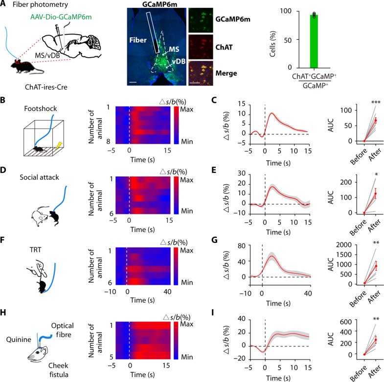
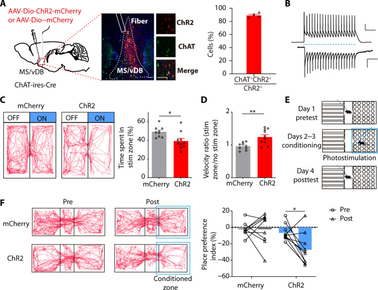
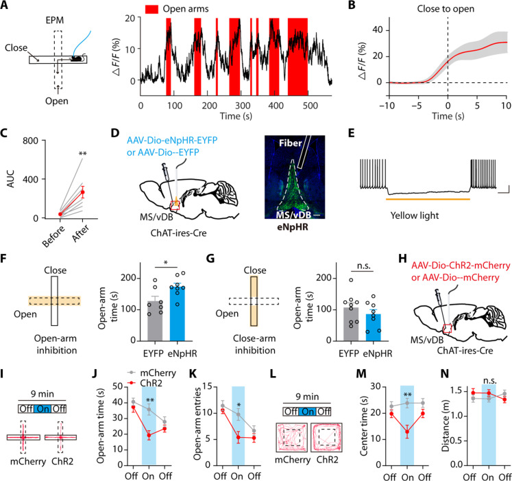
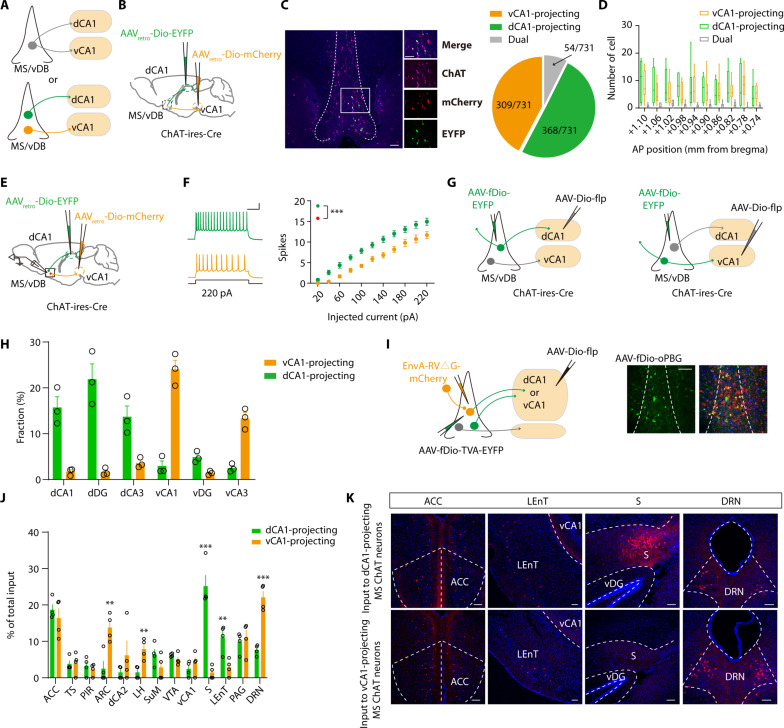
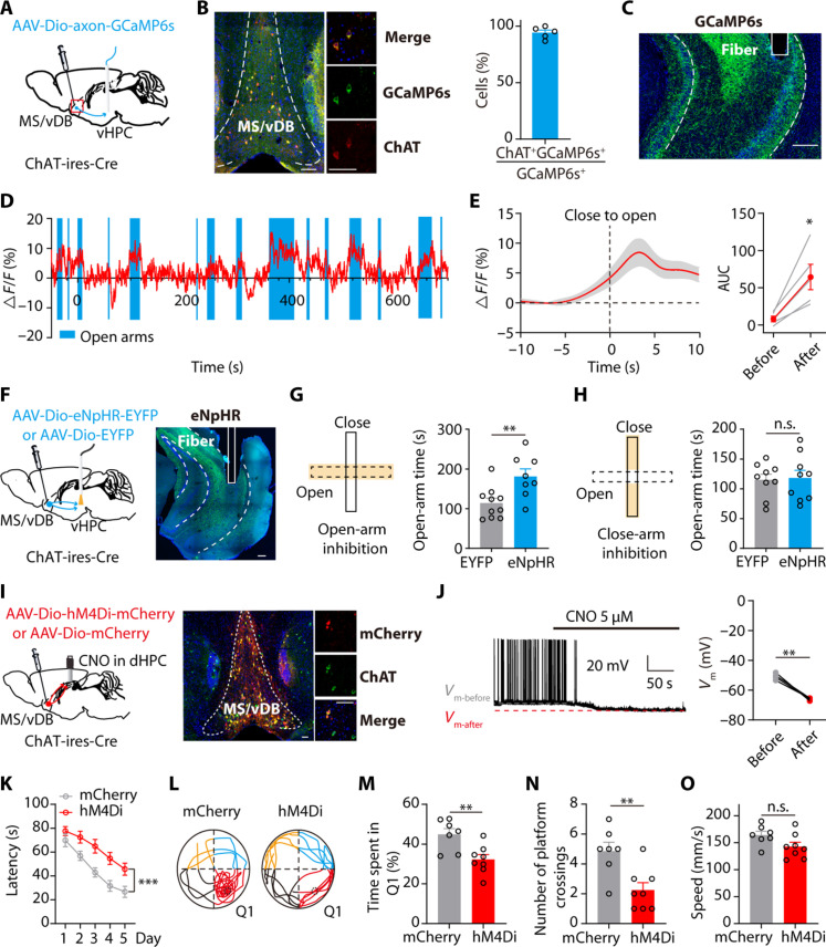
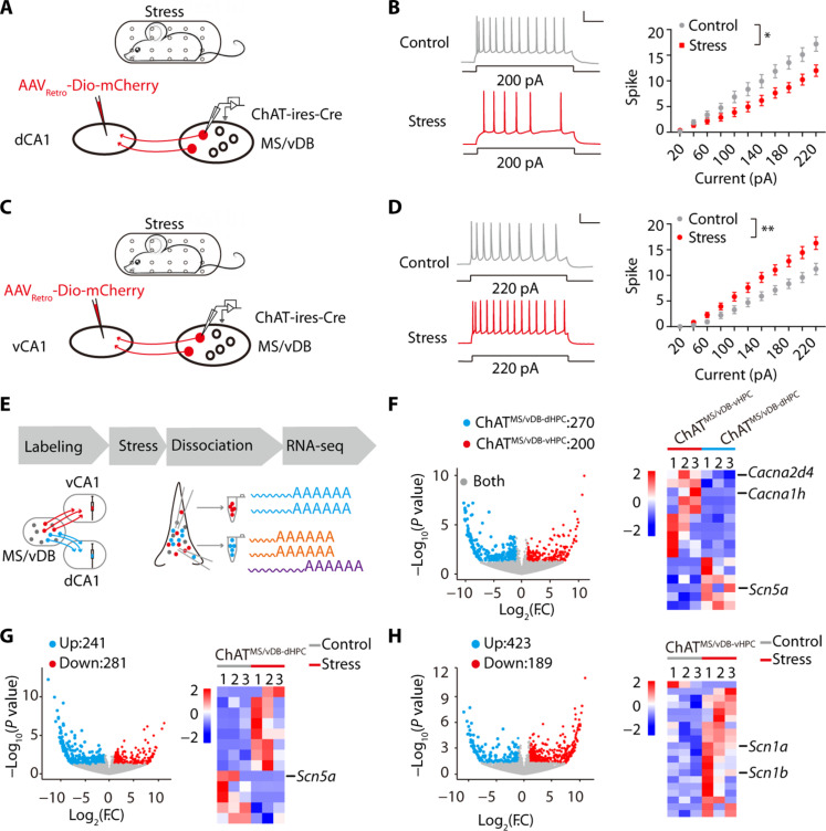
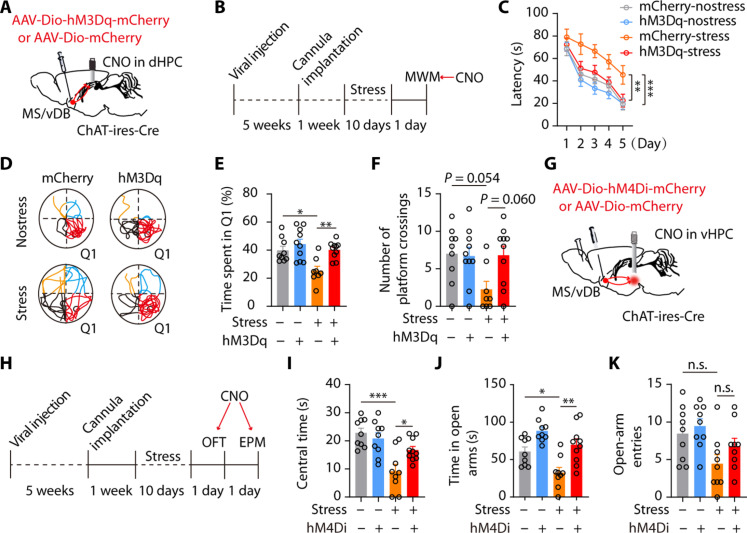
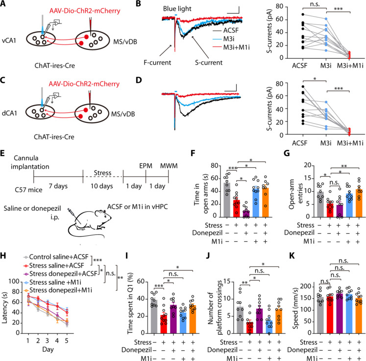
Patients suffering from chronic stress develop numerous symptoms, including emotional and cognitive deficits. The precise circuit mechanisms underlying different symptoms remain poorly understood. We identified two distinct basal forebrain cholinergic subpopulations in mice projecting to the dorsal hippocampus (dHPC) or ventral hippocampus (vHPC), which exhibited distinct input organizations, electrophysiological characteristics, transcriptomics, and responses to positive and negative valences of stimuli and were critical for cognitive and emotional modulation, respectively. Moreover, chronic stress induced elevated anxiety levels and cognitive deficits in mice, accompanied by enhanced vHPC but suppressed dHPC cholinergic projections. Chemogenetic activation of dHPC or inhibition of vHPC cholinergic projections alleviated stress-induced aberrant behaviors. Furthermore, we identified that the acetylcholinesterase inhibitor donepezil combined with blockade of muscarinic receptor 1-type muscarinic acetylcholine receptors in the vHPC rescued both stress-induced phenotypes. These data illuminated distinct septo-hippocampal cholinergic circuits mediated specific symptoms independently under stress, which may provide promising strategies for circuit-based treating of stress-related psychiatric disorders.


Figures

Fig. 1.
Fig. 1.. Aversive stimuli elicit MS/vDB ChAT neuronal activity.
(A) (Left) Fiber photometry setup. (Middle) GCaMP6m expression and optical fiber tract in the MS/vDB. Scale bar, 200 μm. (Further right) Overlap between GCaMP6m-expressing cells (green) and anti–ChAT-positive cells (red). Scale bar, 50 μm. (Right) Percentage of GCaMP6m+ neurons coexpressing ChAT. (B) (Left) Schematic for footshock. (Right) Heatmap of Ca2+ transients across animals aligned to the start of footshock. (C) (Left) Plot of Ca2+ transients across animals aligned to the start of footshock. (Right) Change in calcium signals. n = 8 mice, AUC, area under the curve. (D) (Left) Schematic for social attack. (Right) Heatmap plot of Ca2+ transients across animals aligned to the start of attack. (E) (Left) Plot of Ca2+ transients across animals aligned to the start of attack. (Right) Change in calcium signals. n = 6 mice. (F) (Left) Schematic for TRT application. (Right) Heatmap plot of Ca2+ transients across animals aligned to the start of the TRT. (G) (Left) Plot of Ca2+ transients across animals aligned to the start of the TRT. (Right) Change in calcium signals. n = 6 mice. (H) (Left) Schematic of intraoral quinine infusion. (Right) Heatmap of Ca2+ transients across animals aligned to the start of quinine infusion. (I) (Left) Plot of Ca2+ transients across animals aligned to the start of quinine infusion. (Right) Change in calcium signals. n = 6 mice. Paired t test. *P < 0.05; **P < 0.01; ***P < 0.001. Data are presented as the means ± SEM.
Fig. 2.
Fig. 2.. Activation of MS/vDB ChAT neurons induces place avoidance.
(A) (Left) Schematic of viral injection and optical fiber implantation site. (Middle) ChR2 expression in ChAT neurons and the optical fiber tract above the MS/vDB. Scale bar, 100 μm. (Further right) Overlap between ChR2-expressing cells (red) and anti–ChAT-positive cells (green). Scale bar, 20 μm. (Right) Percentage of GCaMP6m+ neurons coexpressing ChAT (n = 4 mice). (B) Response of a ChR2 cell to a train of light pulses (20 Hz, 10-ms pulse width; blue dashed line) for 1 s in current clamp (top; scale bar, 20 mV, 200 ms) and voltage clamp (bottom; scale bar, 200 pA, 200 ms) mode. (C) (Left) Representative locomotor traces of mCherry and ChR2 mice in an RTPA. (Right) Time spent in the stimuli zone of mice. mCherry, n = 9; ChR2, n = 10; unpaired t test. (D) Velocity ratio of mice. mCherry, n = 9; ChR2, n = 10; unpaired t test. (E) Schematic for the conditioned place preference experiment. (F) (Left) Movement tracking traces for the mCherry mouse and ChR2 mouse in pretest and posttest. (Right) Individual place preference indices (%) of mice before and after conditioning. mCherry, n = 9; ChR2, n = 9; two-way ANOVA with Holm-Sidak post hoc analysis. *P < 0.05; **P < 0.01. Data are presented as the means ± SEM.
Fig. 3.
Fig. 3.. The MS/vDB ChAT neurons process and modulate anxiety-like behaviors.
(A) (Left) Ca2+ recording during the EPM test. (Right) Example of MS/vDB ChAT neuronal activity within the EPM. (B) Plot of Ca2+ transients aligned to the start of open-arm entry. (C) Change in Ca2+ signals. n = 8. (D) (Left) Virus injection and fiber implantation for optogenetic inhibition of MS/vDB ChAT neurons. (Right) Representative image illustrating eNpHR-expressing cells and the fiber tract in the MS/vDB. Scale bar, 200 μm. (E) Response of an eNpHR cell to yellow light illumination under 30 pA of current injection. (F and G) The laser was triggered on when mice entered the open or closed arms only. Open-arm silencing increased the time spent exploring the open arms [(F) EYFP, n = 7; eNpHR, n = 8]. Silencing in the closed arms had no effect [(G) EYFP, n = 9; eNpHR, n = 8]. (H) Schematic of viral expression and implantation site. (I) Representative tracks during the illumination epoch within the EPM for ChR2 mouse and mCherry mouse. (J and K) Light activation of MS/vDB ChAT neurons resulted in a decrease in the duration mice spent exploring the open arms [(J) mCherry, n = 9; ChR2, n = 10] and the open-arm entries (K). (L) Representative tracks during the illumination epoch within the OFT for ChR2 mouse and mCherry mouse. (M) Time spent in exploring the center in the OFT. (N) Distance traveled in the OFT. Paired t test for (C); unpaired t test for (F) and (G); two-way, repeated-measures ANOVA with Bonferroni’s post hoc analysis for (J), (K), (M), and (N). *P < 0.05; **P < 0.01. Data are presented as the means ± SEM. n.s., not significant.
Fig. 4.
Fig. 4.. Distinct subpopulations of MS/vDB ChAT neurons project to the dCA1 and vCA1.
(A) Two possible projection patterns. (B) Dual retrovirus strategy used for labeling dCA1-projecting or vCA1-projecting MS/vDB ChAT neurons. (C) (Left) EYFP and mCherry expression in MS/vDB ChAT neurons (purple). Scale bar, 100 μm. (Right) Proportion of cells classified as projecting to the dCA1, vCA1, or both (731 cells from four mice). (D) Number of dCA1-projecting and vCA1-projecting ChAT neurons throughout the anterior-posterior (AP) extent of the MS/vDB. (E) Ex vivo recording of dCA1-projecting or vCA1-projecting MS/vDB ChAT neurons. (F) Representative traces (left) and injected current-evoked firing relationship (right) of dCA1-projecting or vCA1-projecting MS/vDB ChAT neurons. Scale bar, 100 ms, 20 mV. Two-way, repeated-measures ANOVA with Bonferroni’s post hoc analysis. (G) Viral strategy used for tracing the output of ChATMS/vDB-dCA1 or ChATMS/vDB-vCA1 neurons. (H) Fiber quantitation of dCA1-projecting or vCA1-projecting MS/vDB ChAT neurons. (I) (Left) Strategy to map monosynaptic inputs to either ChATMS/vDB-dCA1 or ChATMS/vDB-vCA1 neurons. (Right) Starter cell localization of AAV-fDio-EYFP-TVA, AAV-fDio-oPBG, and EnvA-RV△G-mCherry (yellow arrows). Scale bar, 100 μm. (J) Whole-brain quantitation of inputs to ChATMS/vDB-dCA1 and ChATMS/vDB-vCA1 neurons. Percentage of total cells in a given brain area relative to the total number of brain-wide inputs. n = 4 for each condition, unpaired t test. (K) Representative images of inputs in select brain areas. ACC, anterior cingulate cortex. Scale bars, 100 μm. **P < 0.01; ***P < 0.001. Data are presented as the means ± SEM.
Fig. 5.
Fig. 5.. MS/vDB cholinergic input to the vHPC and the dHPC separately modulates the anxiety-like behavior and spatial learning.
(A) Recording of the MS/vDB-vHPC cholinergic projections. (B) (Left) GCaMP6s expression in the MS/vDB. Scale bar, 100 μm. (Middle) Overlap between GCaMP6s-expressing cells (green) and anti–ChAT-positive cells (red). Scale bar, 20 μm. (Right) Percentage of GCaMP6s+ neurons coexpressing ChAT (n = 5 mice). (C) Placement of fiber optics. Scale bar, 200 μm. (D) Example of vCA1 cholinergic fiber activity. (E) Plot of Ca2+ transients across animals aligned to the start of open-arm entry and the change in Ca2+ signals. n = 8. (F) Optogenetic silencing of MS/vDB-vHPC cholinergic projections. Scale bar, 200 μm. (G and H) Open-arm silencing increased the time spent exploring the open arms [(G) EYFP, n = 10; eNpHR, n = 8]; silencing in the closed arms had no effect [(H) EYFP, n = 9; eNpHR, n = 9]. (I) Chemogenetic inhibition of the MS/vDB-dHPC cholinergic projections [hM4Di (red) and ChAT neurons (green)]. Scale bar, 100 μm. (J) Representative trace and summarized data showing the inhibition of hM4Di cells after 5 μM CNO delivery. n = 4. (K) Escape latency during acquisition training. mCherry, n = 7; hM4Di, n = 8. (L) Representative swimming traces in the probe trial. (M) Time spent in Q1. (N) Number of platform crossings. (O) Speed in the MWM. Paired t test for (E) and (J); unpaired t test for (G), (H), (M), (N), and (O); two-way, repeated-measures ANOVA for (K). *P < 0.05; **P < 0.01; ***P < 0.001. Data are presented as the means ± SEM.
Fig. 6.
Fig. 6.. ChATMS/vDB-dHPC and ChATMS/vDB-vHPC neurons exhibit distinct electrophysiological and transcriptional adaptations to chronic restraint stress.
(A) Schematic of chronic restraint stress and recording of ChATMS/vDB-dHPC neurons. (B) (Left) Representative traces. Scale bar, 100 ms, 20 mV. (Right) Injected current-evoked firing relationship of ChATMS/vDB-dHPC neurons. Control, n = 15; stress, n = 15. (C) Schematic of chronic restraint stress and recording of ChATMS/vDB-vHPC neurons. (D) (Left) Representative traces. Scale bar, 100 ms, 20 mV. (Right) Injected current-evoked firing relationship of ChATMS/vDB-vHPC neurons. Control, n = 14; stress, n = 15. (E) Strategy of neuronal type–specific RNA-seq. (F) (Left) Scatterplots showing the genes enriched in ChATMS/vDB-vHPC (red) versus ChATMS/vDB-dHPC (green) neurons and both (gray). (Right) Heatmap showing ChATMS/vDB-dHPC and ChATMS/vDB-vHPC neuron-enriched ion channel-related genes. F.C, fold change. (G) (Left) Scatterplots showing the genes down-regulated (red) versus up-regulated (green) in ChATMS/vDB-dHPC neurons after chronic stress. (Right) Heatmap showing ion channel-related genes in ChATMS/vDB-dHPC neurons with a notable change after chronic stress. (H) (Left) Scatterplots showing the genes down-regulated (red) versus up-regulated (green) in ChATMS/vDB-vHPC neurons after chronic stress. (Right) Heatmap showing ion channel-related genes in ChATMS/vDB-vHPC neurons with a notable change after chronic stress. Two-way, repeated-measures ANOVA for (B) and (D). *P < 0.05; **P < 0.01. Data are presented as the means ± SEM.
Fig. 7.
Fig. 7.. Manipulating different septo-hippocampal cholinergic pathways can rescue memory deficits and anxiety-like behaviors induced by chronic stress.
(A) Chemogenetic activation of the MS/vDB-dHPC cholinergic projections. (B) Timeline of experiments. (C) Escape latency during acquisition training. mCherry-nostress, n = 9; hM3Dq-nostress, n = 10; mCherry-stress, n = 8; hM3Dq-stress, n = 10. (D) Representative traces of swimming paths in the probe trial. (E) Time spent in Q1. (F) Number of platform crossings. (G) Schematic for injection of hM4Di and control virus in MS/vDB and CNO delivery in the vHPC. (H) Timeline of experiments. (I) Time spent in the center in stressed mice. mCherry-nostress, n = 9; hM4Di-nostress, n = 9; mCherry-stress, n = 9; hM4Di-stress n = 10. (J) Time spent in the open arms. (K) Open-arm entries. Two-way, repeated-measures ANOVA with Tukey’s multiple comparisons test (C) and one-way ANOVA with Tukey’s multiple comparisons test [(E), (F), and (I) to (K)]. *P < 0.05; **P < 0.01; ***P < 0.001. Data are presented as the means ± SEM.
Fig. 8.
Fig. 8.. AChEIs combined with blockade of the M1 receptor ameliorate both cognitive deficits and anxiety-like behaviors in stressed mice.
(A and C) Schematic of viral injection, the location of light stimuli, and the recording configuration in acute slices. (B) Representative traces (left) and quantification (right) show light-evoked muscarinic inward currents were suppressed by application of the M3 inhibitor DAU5884 (M3i; 5 μM) but not the M1 inhibitor VU0255035 (M1i; 5 μM). Scale bar, 100 ms, 20 pA (n = 12 neurons from four mice). (D) Representative traces (left) and quantification (right) show muscarinic inward currents were suppressed by application of M3i or M1i. Scale bar, 100 ms, 20 pA (n = 11 neurons from four mice). (E) Timeline of experiments and scheme of drug delivery strategy. (F and G) Time spent in the open arms [(F) n = 8 to 10) and open-arm entries (G). (H) Donepezil injection combined with intra-vHPC delivery of M1i in stress mice during the training session reversed the impairment of spatial learning in the MWM. (I to K) Donepezil injection combined with intra-vHPC delivery of M1i during the training session in stressed mice increased the time spent in Q1 (I) and the number of platform crossings (J) with no effect on speed (K). One-way ANOVA with Tukey’s multiple comparison test for (B) and (D) and two-way ANOVA with Bonferroni’s multiple comparison test for (F), (G), (I), (J), and (K). *P < 0.05; **P < 0.01; ***P < 0.001. Data are presented as the means ± SEM.

References

    1. McEwen B. S., Bowles N. P., Gray J. D., Hill M. N., Hunter R. G., Karatsoreos I. N., Nasca C., Mechanisms of stress in the brain. Nat. Neurosci. 18, 1353–1363 (2015). - PMC - PubMed
    1. Fenster R. J., Lebois L. A. M., Ressler K. J., Suh J., Brain circuit dysfunction in post-traumatic stress disorder: From mouse to man. Nat. Rev. Neurosci. 19, 535–551 (2018). - PMC - PubMed
    1. Schwabe L., Hermans E. J., Joëls M., Roozendaal B., Mechanisms of memory under stress. Neuron 110, 1450–1467 (2022). - PubMed
    1. de Quervain D., Schwabe L., Roozendaal B., Stress, glucocorticoids and memory: Implications for treating fear-related disorders. Nat. Rev. Neurosci. 18, 7–19 (2017). - PubMed
    1. Ballinger E. C., Ananth M., Talmage D. A., Role L. W., Basal forebrain cholinergic circuits and signaling in cognition and cognitive decline. Neuron 91, 1199–1218 (2016). - PMC - PubMed

Publication types