

Cystic lymphangioma of the head and tail of the pancreas in a middle-aged Iranian woman: A case report
- PMID: 39515211
- PMCID: PMC11570977
- DOI: 10.1016/j.ijscr.2024.110453
Cystic lymphangioma of the head and tail of the pancreas in a middle-aged Iranian woman: A case report
Abstract
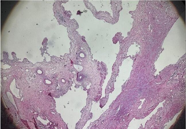
Introduction: Lymphangioma is a benign neoplasm that involves the lymphatic vessels in multiple anatomical regions, including the pancreas, where it is exceptionally rare, with a prevalence of only 0.2 %. These tumors, often found in children, are uncommon in adults and may present with symptoms like abdominal pain, distention, and nausea. Many cases are asymptomatic and discovered incidentally during imaging studies. A definitive diagnosis is achieved through pathological examination following a biopsy. This report details a rare instance of pancreatic lymphangioma in a 58-year-old Iranian woman experiencing acute abdominal symptoms.
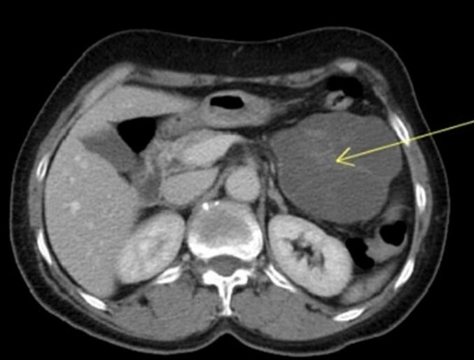
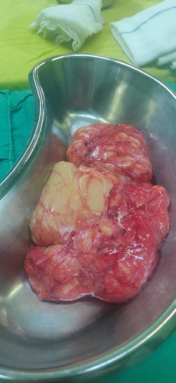
Case presentation: A 58-year-old woman presented with acute abdominal pain, nausea, and vomiting, ultimately diagnosed with pancreatic lymphangioma. She underwent exploratory laparotomy, resulting in the successful excision of a cystic mass from the pancreas. Histopathological examination confirmed the diagnosis. This case emphasizes the necessity of considering pancreatic lymphangioma in the differential diagnosis of acute abdominal pain in adults, where it is less frequently encountered. Ultrasonography and CT scans played pivotal roles in the diagnosis, while surgical excision remains the gold standard for both diagnosis and treatment. Physical examination revealed left upper quadrant tenderness and a large palpable mass. Laboratory tests ruled out acute pancreatitis or myocardial infarction.
Discussion: Pancreatic lymphangiomas are exceedingly rare and often asymptomatic. When symptomatic, they can present with acute abdominal pain and other gastrointestinal disturbances. The etiology may relate to congenital malformations or inflammatory processes affecting lymphatic vessels. The case highlights the necessity of including pancreatic lymphangioma in differential diagnoses for abdominal pain in adults, despite its rarity. While ultrasonography remains a primary diagnostic modality, CT and MRI are invaluable for providing comprehensive evaluations of cystic lesions. Magnetic resonance cholangiopancreatography (MRCP) can assist in distinguishing lymphangiomas from other cystic lesions, although it may not always yield definitive results without further invasive procedures like endoscopic ultrasound-guided fine needle aspiration (EUS-FNA).
Conclusion: This case contributes to the limited literature on pancreatic lymphangioma in adults and highlights the need for further research to develop standardized medical approaches for managing this rare condition. The findings suggest that while pancreatic lymphangiomas are benign, they should be included in differential diagnoses for pancreatic cystic lesions, given their potential for causing significant symptoms.
Keywords: Acute abdominal pain; Case report; Cystic lymphangioma; Pancreas; Pancreatic cyst.
Copyright © 2024 The Authors. Published by Elsevier Ltd.. All rights reserved.
Conflict of interest statement
Declaration of competing interest The authors declare no competing interests.
Figures
Similar articles
-
Recurrence of a pancreatic cystic lymphangioma after diagnosis and complete drainage by endoscopic ultrasound with fine-needle aspiration.JOP. 2013 May 10;14(3):280-2. doi: 10.6092/1590-8577/1347. JOP. 2013. PMID: 23669480
-
Symptomatic diffuse cystic lymphangioma of the spleen in an adult: A case report.Int J Surg Case Rep. 2025 Jan;126:110669. doi: 10.1016/j.ijscr.2024.110669. Epub 2024 Nov 28. Int J Surg Case Rep. 2025. PMID: 39612903 Free PMC article.
-
Cystic lymphangioma of the pancreas: a hard diagnostic challenge between pancreatic cystic lesions-review of recent literature.Gland Surg. 2018 Oct;7(5):487-492. doi: 10.21037/gs.2018.04.02. Gland Surg. 2018. PMID: 30505770 Free PMC article.
-
Pancreatic Lymphangioma: A Case Report and Literature Review.J Comput Assist Tomogr. 2019 Mar/Apr;43(2):242-244. doi: 10.1097/RCT.0000000000000818. J Comput Assist Tomogr. 2019. PMID: 30371621 Review.
-
[Perirenal cystic lymphangioma in an adult: a case report and literature review].Beijing Da Xue Xue Bao Yi Xue Ban. 2017 Aug 18;49(4):730-732. Beijing Da Xue Xue Bao Yi Xue Ban. 2017. PMID: 28816297 Review. Chinese.
References
-
- Cystic lymphangioma of the pancreas Kostov, D. et al. HPB, Volume 21, S687, doi:10.1016/j.hpb.2019.10.528. - DOI
Publication types
LinkOut - more resources
Full Text Sources
