. 2024 Dec 10;19(12):1690-1705.
doi: 10.1016/j.stemcr.2024.10.004. Epub 2024 Nov 7.

Derivation of transplantable human thyroid follicular epithelial cells from induced pluripotent stem cells

Affiliations

Derivation of transplantable human thyroid follicular epithelial cells from induced pluripotent stem cells

Hendrik J Undeutsch et al. Stem Cell Reports. .

Abstract

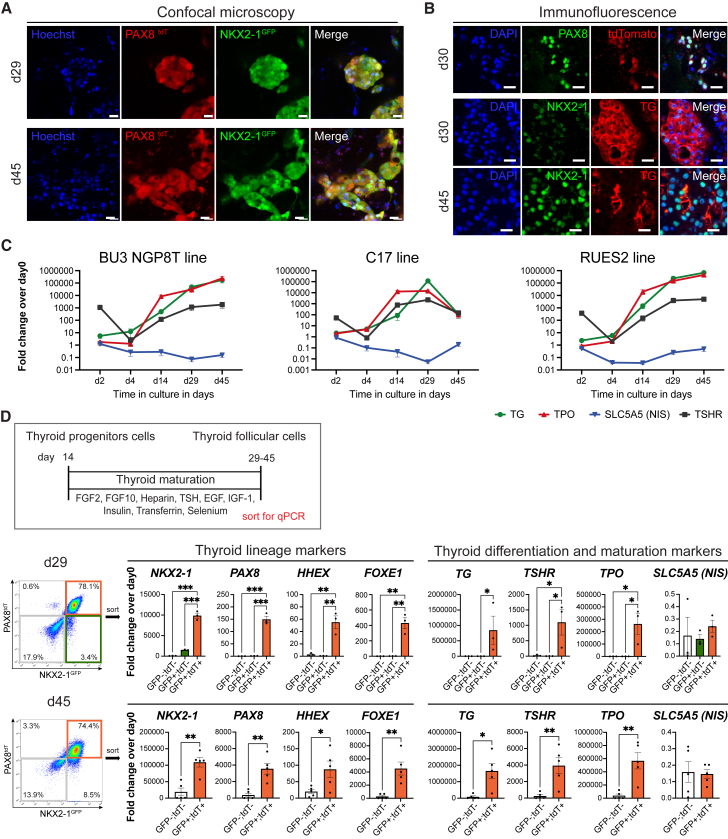
The production of mature functioning thyroid follicular cells (TFCs) from human induced pluripotent stem cells (iPSCs) is critical for potential novel therapeutic approaches to post-surgical and congenital hypothyroidism. To accomplish this, we developed a novel human iPSC line that expresses fluorophores targeted to the NKX2-1 and PAX8 loci, allowing for the identification and purification of cells destined to become TFCs. Optimizing a sequence of defined, serum-free media to promote stepwise developmental directed differentiation, we found that bone morphogenic protein 4 (BMP4) and fibroblast growth factor 2 (FGF2) stimulated lineage specification into TFCs from multiple iPSC lines. Single-cell RNA sequencing demonstrated that BMP4 withdrawal after lineage specification promoted TFC maturation, with mature TFCs representing the majority of cells present within 1 month. After xenotransplantation into athyreotic immunodeficient mice, engrafted cells exhibited thyroid follicular organization with thyroglobulin protein detected in the lumens of NKX2-1-positive follicles. While our iPSC-derived TFCs presented durable expression of thyroid-specific proteins, they were unable to rescue hypothyroidism in vivo.

Keywords: directed differentiation; human; organoids; pluripotent stem cells; thyroid.


Conflict of interest statement

Declaration of interests The authors declare no competing interests.

Figures

None
Graphical abstract
Figure 1
Figure 1
Generation of NKX2-1GFP;PAX8tdTomato reporter hiPSC line (A) Gene editing constructs to target a GFP reporter to the human NKX2-1 locus (Hawkins et al., 2017) and a tdTomato reporter to the human PAX8 locus to generate a NKX2-1GFP;PAX8tdT double reporter hiPSC line. (B) Schematic overview of directed differentiation of hiPSC into NKX2-1GFP+;PAX8tdT+ thyroid progenitor cells. (C) Efficiency of definitive endoderm induction (DEI) assessed by FACS for CKIT; CXCR4 co-expression on day 2 and 3 of DEI (upper) and by FACS for NKX2-1GFP;PAX8tdT expression on day 14 (lower), comparing outgrowth following 2 and 3 days of DEI (n = 3, unpaired t test). (D) Gene expression for endodermal markers at day 2, 4, and 6 of differentiation with 2 days of DEI (n = 3, one-way ANOVA with Tukey’s multiple comparison). (E) Gene expression of NKX2-1 and PAX8 of sorted cells at day 12 (n = 3, one-way ANOVA with Dunnett’s post hoc test). Data shown in (C–E) are of 3 independent experiments. p < 0.05; ∗∗p < 0.01; ∗∗∗p < 0.001; error bars represent standard error of mean (SEM).
Figure 2
Figure 2
NKX2-1GFP+;PAX8tdT+ cells are early thyroid follicular progenitors by day 14, uniquely co-expressing thyroid lineage-selective transcription factors (A) Schematic timeline for thyroid development in mice and humans. (B) Immunofluorescent staining at day 14 for NKX2-1 and PAX8 in the BU3 NGP8T line (scale bar, 20 μm). (C) Immunofluorescent staining for NKX2-1 and GFP expression (upper) and PAX8 and tdTomato (tdT) expression (lower) in BU3 NGP8T cells at day 14 (scale bar, 20 μm). (D) Confocal microscopy at day 14 detecting endogenous NKX2-1GFP and PAX8tdT expression in thyroid progenitors derived from BU3 NGP8T cells (top) and NKX2-1GFP expression in the C17 cell line (lower, scale bar, 50 μm). (E) Gene expression profiling of BU3 NGP8T line after sorting for NKX2-1GFP and PAX8tdT on day 14 of thyroid specification (n = 3 samples from independent experiments; one-way ANOVA with Dunnett’s multiple comparison test; p < 0.05; ∗∗p < 0.01; ∗∗∗p < 0.001; error bars represent SEM).
Figure 3
Figure 3
Extended BMP signaling prevents the maturation of thyroid progenitors toward functional thyroid cells (A) Schematic overview of experimental setup for scRNA-seq comparing thyroid progenitor cells (day 12) to thyroid follicular cells (TFCs) at day 29 cultured in presence (with BMP) or absence (w/o BMP) of BMP4 after day 14. (B) Heatmap with top 50 differentially expressed genes ranked by p value. (C) scRNA-seq of day 12 thyroid progenitor cells and day 29 TFCs cultured in maturation media or maturation media + BMP4. UMAP (left) displays the distribution-based time point and culture conditions used to treat cells. Louvain clustering (resolution = 0.15) identifies 7 clusters (right). (D) UMAP of thyroid lineage markers (upper), thyroid differentiation, and maturation markers (lower). (E) UMAP of the reporter genes targeted to the NKX2-1 and PAX8 locus. (F) Bubble plot for thyroid-specific genes for the different time points and treatment. (G) Violin plots for thyroid lineage, differentiation, and maturation marker signatures by time point and treatment (one-way ANOVA, ∗∗∗p < 0.001). (H) Distribution of cells based on cycle phases by time point and culture condition.
Figure 4
Figure 4
Differentiation and maturation of NKX2-1GFP+;PAX8tdT+ thyroid progenitor cells to TFCs is time dependent (A) Confocal imaging of BU3 NGP8T line at 29 (upper) and 45 days (lower) of differentiation maintains NKX2-1GFP and PAX8tdT reporter expression (scale bar, 20 μm). (B) Immunofluorescent staining for PAX8 and the tdT reporter in day 30 organoids (upper) and for TG protein expression in NKX2-1-positive organoids at day 30 (middle) and day 45 (lower; scale bar = 20 μm). (C) Time-dependent gene expression analysis for thyroid differentiation and maturation at day 0, 2, 4, 14, 29, and 45 of differentiation in BU3 NGP8T (left; n = 3–4 samples from 4 independent experiment), C17 (middle; n = 3 replicates from 1 experiment), and RUES2 (right; n = 4–6 from 6 independent experiments). (D) Schematic overview of thyroid maturation protocol (top). Gene expression profiling for thyroid lineage, differentiation, and maturation markers in subpopulations of BU3 NGP8T cells sorted for cells expressing no reporter, NKX2-1GFP+ single-positive cells, and NKX2-1GFP+;PAX8tdT+ double-positive cells at day 29 (middle; n = 3 samples from 3 independent experiments, one-way ANOVA with Kruskal-Wallis post hoc test; p < 0.05; ∗∗p < 0.01; ∗∗∗p < 0.001) and NKX2-1GFP−;PAX8tdT− double-negative and NKX2-1GFP+;PAX8tdT+ double-positive cells at day 45 (lower; n = 5 from 5 independent experiments, unpaired t test; p < 0.05; ∗∗p < 0.01). Error bars in (C) and (D) represent SEM.
Figure 5
Figure 5
hiPSC-derived organoids transplanted under renal capsule of hypothyroid, immunocompromised mice form thyroid follicles but did not restore normal thyroid hormone status (A) Schematic overview of subrenal capsule graft experiment. (B) Total T4 (upper) and TSH (lower) of radioiodine-ablated mice prior to surgery (left side) confirmed hypothyroidism following ablation. T4 and TSH levels did not normalize up to 12 weeks after transplantation (right side; p value determined by Brown-Forsythe’s one-way ANOVA [T4] and Kruskal-Wallis tests [TSH], n = 2–13 mice per group, samples with n < 3 were excluded from statistical analysis. p < 0.05; ∗∗p < 0.01; ∗∗∗p < 0.001; error bars represent SEM). (C) Representative images of H&E staining of subrenal kidney grafts using undifferentiated (day 0) hiPSC, hiPSC-derived thyroid organoids cultured for 30 and 47 days (scale bar, 100 μm). Selected areas are shown with higher magnification below (scale bar, 50 μm). (D) Immunofluorescence staining of subrenal grafts of day 30 (upper row) and day 47 cells (lower row) for NKX2-1 and TG (scale bar, 50 μm). Merged image for selected areas is shown with higher magnification on the right, together with a second subrenal graft of day 47 organoids (bottom, scale bar, 20 μm).
Figure 6
Figure 6
Vascularization of transplanted organoids and analysis of MAPK inhibition to enhance NIS expression (A) Immunofluorescent staining for hNuMA and isolectin B4 (IB4) of a day 30 kidney graft and (B) for PAX8 and CD31 of a day 47 graft recipient (scale bar, 50 μm). Selected areas are shown with higher magnification as merged overlay and bright-field images on the right, mark lumen of follicular structures (scale bar, 20 μm). (C) Gene expression analysis for the effect of PD98059 (PD) and cAMP/IBMX (CI) treatment for 3, 6, or 9 days during maturation (between day 26 and 44) compared to untreated cells. NKX2-1 (left), TG (middle), and SLC5A5 (right) expression of unsorted cells at day 29 to 44 (n = 6–18 samples from 3 independent experiments, combined from 2 to 6 time points. One-way ANOVA with Kruskal-Wallis post hoc test; ∗∗p < 0.01; error bars represent SEM).

Similar articles

Cited by

References

    1. Afink G.B., Veenboer G., de Randamie J., Keijser R., Meischl C., Niessen H., Ris-Stalpers C. Initial characterization of C16orf89, a novel thyroid-specific gene. Thyroid. 2010;20:811–821. - PubMed
    1. Antonica F., Kasprzyk D.F., Opitz R., Iacovino M., Liao X.H., Dumitrescu A.M., Refetoff S., Peremans K., Manto M., Kyba M., Costagliola S. Generation of functional thyroid from embryonic stem cells. Nature. 2012;491:66–71. - PMC - PubMed
    1. Barker N., van Es J.H., Kuipers J., Kujala P., van den Born M., Cozijnsen M., Haegebarth A., Korving J., Begthel H., Peters P.J., Clevers H. Identification of stem cells in small intestine and colon by marker gene Lgr5. Nature. 2007;449:1003–1007. - PubMed
    1. Bayha E., Jørgensen M.C., Serup P., Grapin-Botton A. Retinoic acid signaling organizes endodermal organ specification along the entire antero-posterior axis. PLoS One. 2009;4 - PMC - PubMed
    1. Bouchard M., Souabni A., Busslinger M. Tissue-specific expression of cre recombinase from the Pax8 locus. Genesis. 2004;38:105–109. - PubMed

Publication types

MeSH terms

LinkOut - more resources