. 2024 Nov;30(11):e70071.
doi: 10.1111/cns.70071.

PTP1B Modulates Carotid Plaque Vulnerability in Atherosclerosis Through Rab5-PDGFRβ-Mediated Endocytosis Disruption and Apoptosis

Affiliations

PTP1B Modulates Carotid Plaque Vulnerability in Atherosclerosis Through Rab5-PDGFRβ-Mediated Endocytosis Disruption and Apoptosis

Xiao Zhang et al. CNS Neurosci Ther. 2024 Nov.

Abstract

Background: Protein tyrosine phosphatase 1B (PTP1B) is a protein tyrosine phosphatase and modulates platelet-derived growth factor (PDGF)/platelet-derived growth factor receptor (PDGFR) signaling in vascular smooth muscle cells (VSMCs) via endocytosis. However, the related molecular pathways that participated in the interaction of endo-lysosome and the trafficking of PDGFR are largely unknown. This study aims to determine the subcellular regulating mechanism of PTP1B to the endo-lysosome degradation of PDGFR in atherosclerotic carotid plaques, thereby offering a potential therapeutic target for the stabilization of carotid plaques.

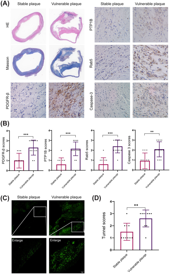
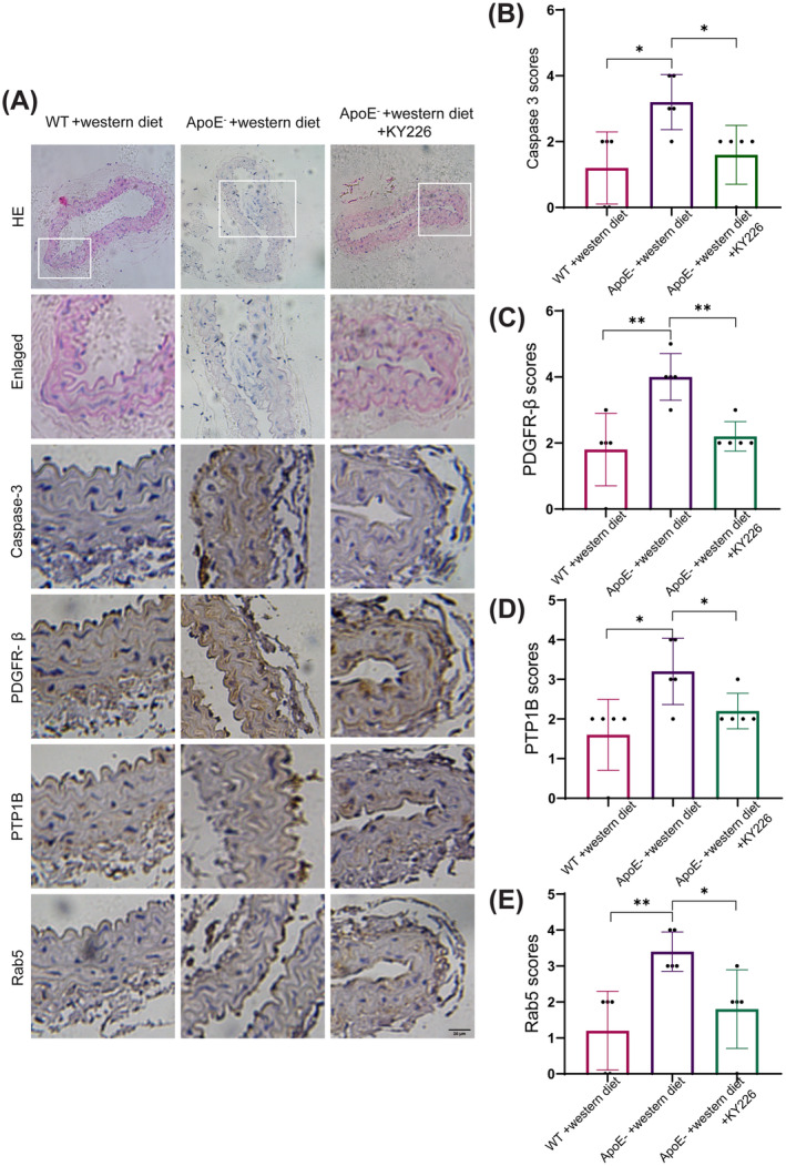
Methods: The immunohistochemical staining technique was employed to assess the expression levels of both PDGFR-β and Caspase 3 in stable and vulnerable carotid plaques. Tunnel staining was utilized to quantify the apoptosis of carotid plaques. Live-cell imaging was employed to observe endocytic motility, while cell apoptosis was evaluated through Propidium Iodide staining. In an in vivo experiment, ApoE-/- mice were administered a PTP1B inhibitor to investigate the impact of PTP1B on atherosclerosis.

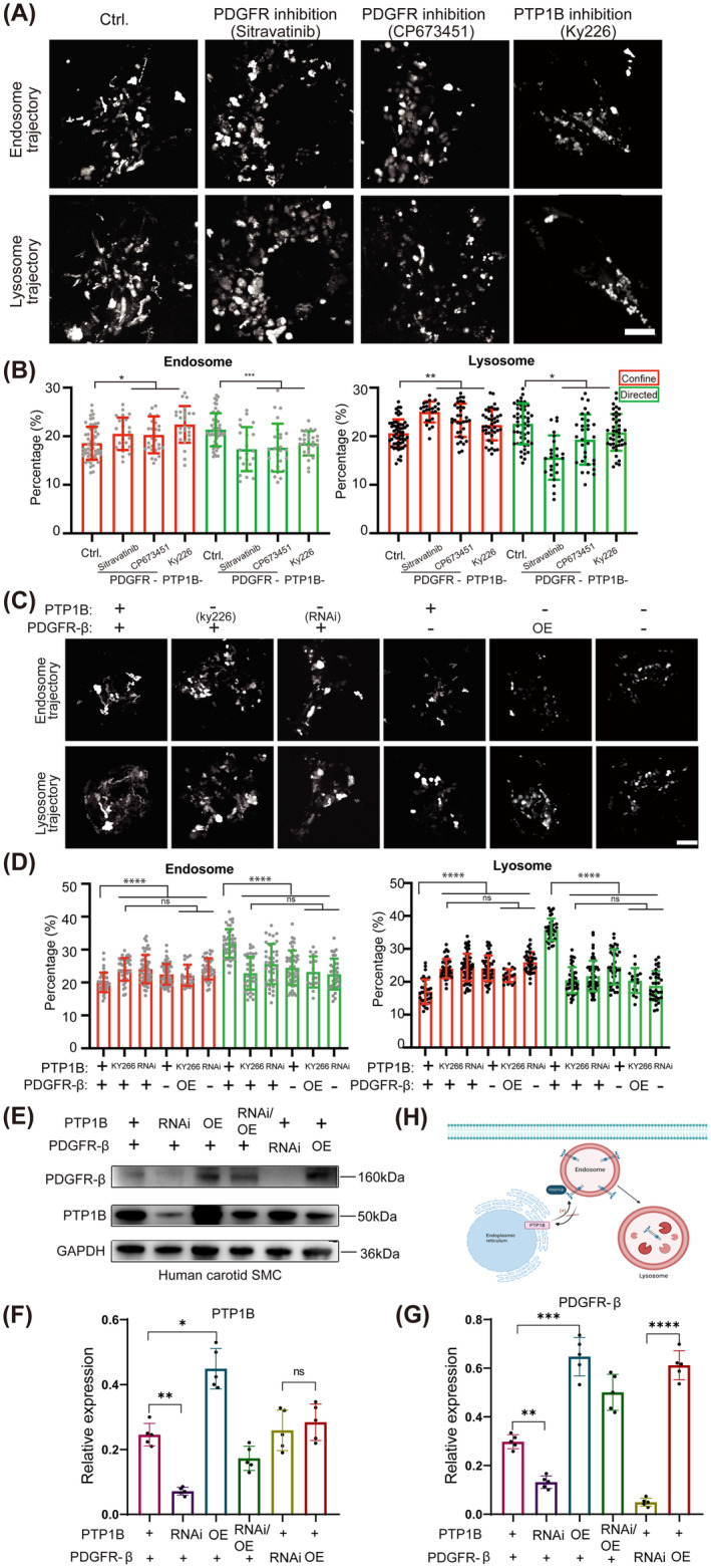
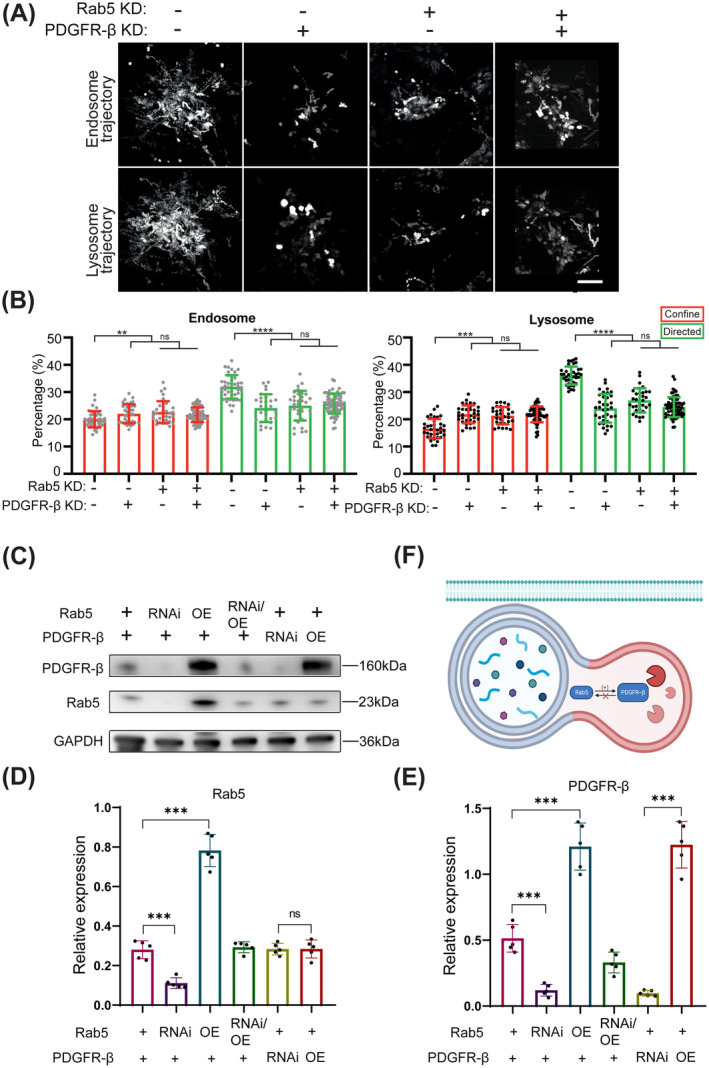
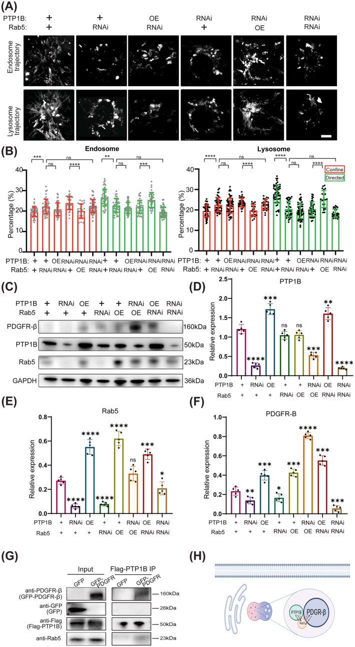
Results: The heightened expression of PDGFR-β correlates with apoptosis in patients with vulnerable carotid plaques. At the subcellular level of VSMCs, PDGFR-β plays a pivotal role in sustaining a balanced endocytosis system motility, regulated by the expression of Rab5, a key regulator of endocytic motility. And PTP1B modulates PDGFR-β signaling via Rab5-mediated endocytosis. Additionally, disrupted endocytic motility influences the interplay between endosomes and lysosomes, which is crucial for controlling PDGFR-β trafficking. Elevated PTP1B expression induces cellular apoptosis and impedes migration and proliferation of carotid VSMCs. Ultimately, mice with PTP1B deficiency exhibit a reduction in atherosclerosis.

Conclusion: Our results illustrate that PTP1B induces disruption in endocytosis and apoptosis of VSMCs through the Rab5-PDGFRβ pathway, suggesting a potential association with the heightened vulnerability of carotid plaques.

Keywords: PTP1B; apoptosis; atherosclerosis; carotid plaque; endocytosis disruption.


Conflict of interest statement

The authors declare no conflicts of interest.

Figures

FIGURE 1
FIGURE 1
The accumulation of PDGFR‐β expression leads to apoptosis in patients with vulnerable carotid plaques. HE, Hematoxylin–Eosin. (A) HE, Masson, and immunohistochemical staining for PDGFR‐β, PTP1B, Rab5, and Caspase‐3 in representative stable plaques and vulnerable carotid plaques. (B) Quantitative analysis of immunohistochemical staining scores for PDGFR‐β, PTP1B, Rab5, and Caspase‐3 in stable and vulnerable carotid plaques. (C) Tunnel staining for representative stable plaques and vulnerable carotid plaques. (D) Quantitative analysis of tunnel staining score for stable and vulnerable carotid plaques. Scar bar 10 μm. **p < 0.01, ***p < 0.001.
FIGURE 2
FIGURE 2
PDGFR is required for sustained and balanced motility of the endocytosis system in vascular smooth muscle cells. Ctrl, Control; OE, Overexpression; RNAi, RNA interference. (A) Endosome and lysosome trajectories of carotid VSMCs with the treatment of PDGFR inhibitors (Sitravatinib and CP673452), and PTP1B inhibitor (KY226). (B) Quantitative analysis of confined and directed modes of endosome and lysosome. (C) Endosome and lysosome trajectories of cos7 cells with the treatments of PDGFR‐β inhibitor and overexpression, and PTP1B inhibitor or RNA knockdown. (D) Quantitative analysis of confined and directed modes of endosome and lysosome. (E) Western blot analyses of PDGFR‐β and PTP1B. (F) The relative level of PTP1B/GAPDH (n = 5). (G) The relative level of PDGFR‐β/GAPDH (n = 5). (H) Schematic diagram of the relationship between PTP1B and PDGFR‐β on endocytosis system. Scar bar 10 μm. *p < 0.05, **p < 0.01, ***p < 0.001, ****p < 0.0001, and ns p > 0.05.
FIGURE 3
FIGURE 3
Loss of PDGFR decreases the expression of Rab5 which is responsible for the endocytic motility. KD, Knockdown; OE, Overexpression; RNAi, RNA interference. (A) Endosome and lysosome trajectories of cos7 cells with the treatments of Rab5 knockdown, PDGFR‐β knockdown, or both knockdowns. (B) Quantitative analysis of confined and directed modes of endosome and lysosome. (C) Western blot analyses of PDGFR‐β and Rab5. (D) The relative level of Rab5/GAPDH (n = 5). (E) The relative level of PDGFR‐β/GAPDH (n = 5). (F) Schematic diagram of the relationship between Rab5 and PDGFR‐β on endocytosis system. Scar bar 10 μm. **p < 0.01, ***p < 0.001, ****p < 0.0001, and ns p > 0.05.
FIGURE 4
FIGURE 4
PTP1B modulates PDGFR‐β signaling via Rab5‐mediated endocytosis. OE, Overexpression; RNAi, RNA interference. (A) Endosome and lysosome trajectories of cos7 cells with the treatments of PTP1B knockdown or overexpression, and Rab5 knockdown or overexpression. (B) Quantitative analysis of confined and directed modes of endosome and lysosome. (C) Western blot analyses of PDGFR‐β, PTP1B, and Rab5. (D) The relative level of PTP1B/GAPDH (n = 5). (E) The relative level of Rab5/GAPDH (n = 5). (F) The relative level of PDGFR‐β/GAPDH (n = 5). (G) Western blotting images of Coimmunoprecipitation to test the interaction among PTP1B, Rab5, and PDGFR‐β. (H) Schematic diagram of relationship among PTP1B, Rab5, and PDGFR‐β on endocytosis system. Scar bar 10 μm. *p < 0.05, **p < 0.01, ***p < 0.001, ****p < 0.0001, and ns p > 0.05.
FIGURE 5
FIGURE 5
Increased PTP1B expression leads to cells' apoptosis, and suppresses carotid SMC migration and proliferation. RNAi: RNA interference. OE, Overexpression; PI, Propidium Iodide. Edu: 5‐ethynyl‐2′‐deoxyuridine. (A) Apoptosis was assessed by PI staining of cos7 cells treated with knockdown or overexpression of PTP1B and Rab5, respectively. Scar bar 20 μm. (B) Quantitative analysis of PI‐positive cells among 20 cells. (C) Western blot analyses of Cleaved Caspase‐3 of cos7 cells treated with knockdown or overexpression of PTP1B and Rab5, respectively. (D) The relative level of Cleaved Caspase‐3/GAPDH (n = 5). (E) Edu staining of carotid SMC treated with knockdown or overexpression of PTP1B. Scar bar 10 μm. (F) Scratch test of carotid SMC treated with knockdown or overexpression of PTP1B. Scar bar 10 μm. (G) Quantitative analysis of Edu‐positive cells. (H) Quantitative analysis of wound width. *p < 0.05, **p < 0.01, ***p < 0.001, ****p < 0.0001, and ns p > 0.05.
FIGURE 6
FIGURE 6
PTP1B deficiency decreases mouse atherosclerosis. HE, Hematoxylin–Eosin; WT, Wide type. (A) HE and immunohistochemical staining for Caspase‐3, PDGFR‐β, PTP1B, and Rab5 in representative mouse carotid arteries from Wildtype +western diet, ApoE + western diet and ApoE + western diet +KY226. (B–E) Quantitative analysis of immunohistochemical staining scores for Caspase‐3, PDGFR‐β, PTP1B, and Rab5 in mouse carotid arteries. *p < 0.05, **p < 0.01.

References

    1. Gao P., Wang T., Wang D., et al., “Effect of Stenting Plus Medical Therapy vs Medical Therapy Alone on Risk of Stroke and Death in Patients With Symptomatic Intracranial Stenosis: The CASSISS Randomized Clinical Trial,” Journal of the American Medical Association 328, no. 6 (2022): 534–542. - PMC - PubMed
    1. Brinjikji W., J. Huston, 3rd , Rabinstein A. A., Kim G. M., Lerman A., and Lanzino G., “Contemporary Carotid Imaging: From Degree of Stenosis to Plaque Vulnerability,” Journal of Neurosurgery 124, no. 1 (2016): 27–42. - PubMed
    1. Chang R. W., Tucker L. Y., Rothenberg K. A., et al., “Incidence of Ischemic Stroke in Patients With Asymptomatic Severe Carotid Stenosis Without Surgical Intervention,” Journal of the American Medical Association 327, no. 20 (2022): 1974–1982. - PMC - PubMed
    1. Bentzon J. F., Otsuka F., Virmani R., and Falk E., “Mechanisms of Plaque Formation and Rupture,” Circulation Research 114, no. 12 (2014): 1852–1866. - PubMed
    1. Liu M. and Gomez D., “Smooth Muscle Cell Phenotypic Diversity,” Arteriosclerosis, Thrombosis, and Vascular Biology 39, no. 9 (2019): 1715–1723. - PMC - PubMed

Publication types

MeSH terms

Substances

LinkOut - more resources