. 2024 Oct 24;16(21):3589.
doi: 10.3390/cancers16213589.

Cannabidiol (CBD) Protects Lung Endothelial Cells from Irradiation-Induced Oxidative Stress and Inflammation In Vitro and In Vivo

Affiliations

Cannabidiol (CBD) Protects Lung Endothelial Cells from Irradiation-Induced Oxidative Stress and Inflammation In Vitro and In Vivo

Lisa Bauer et al. Cancers (Basel). .

Abstract

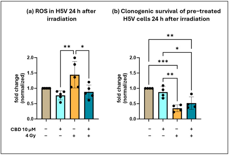
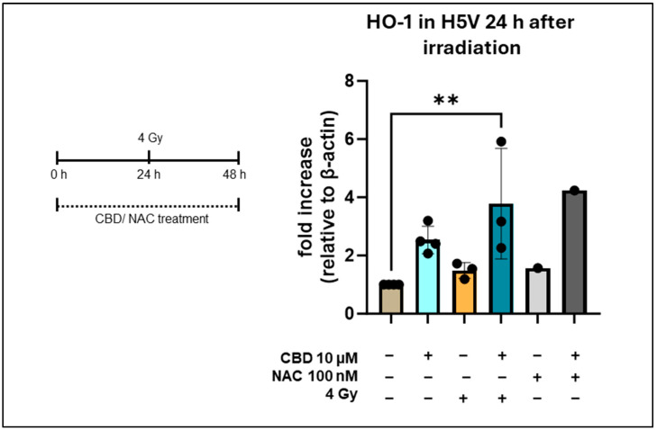
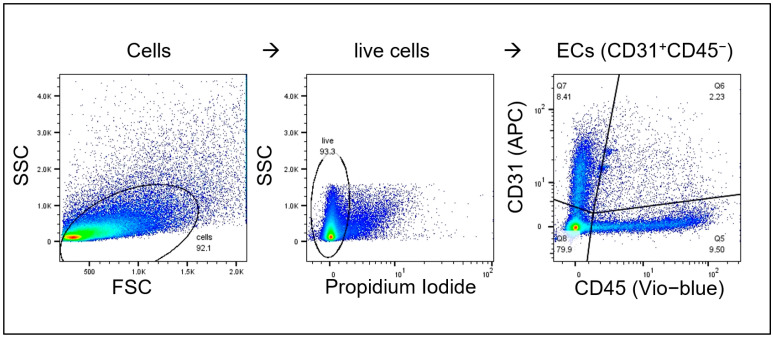
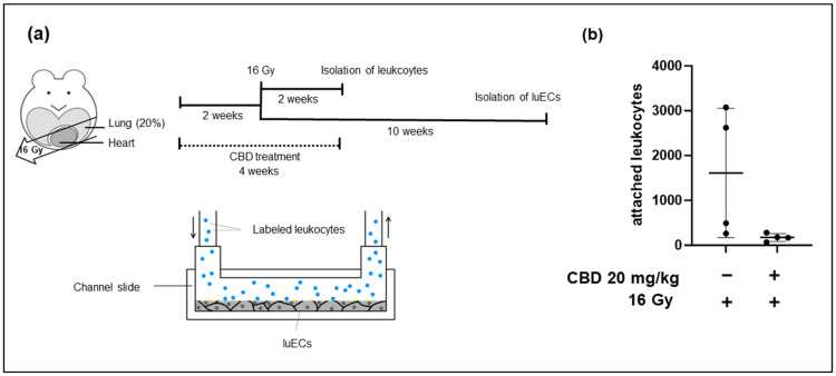
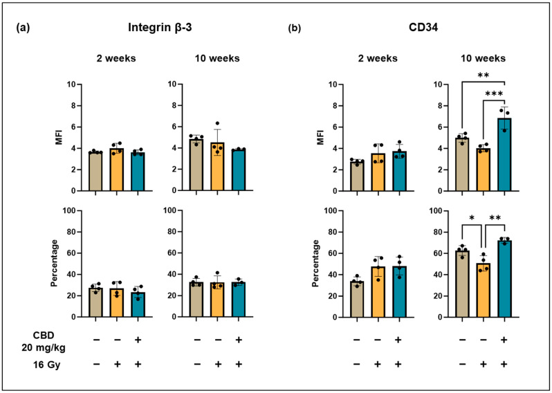
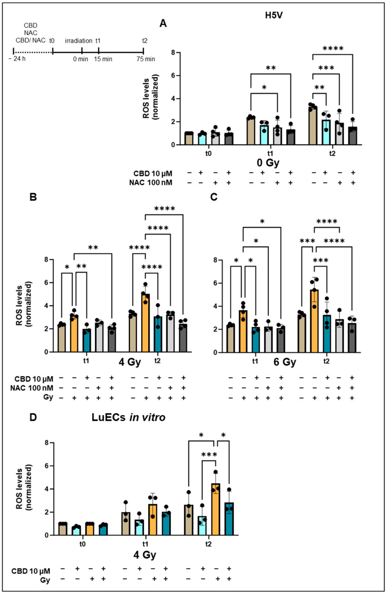
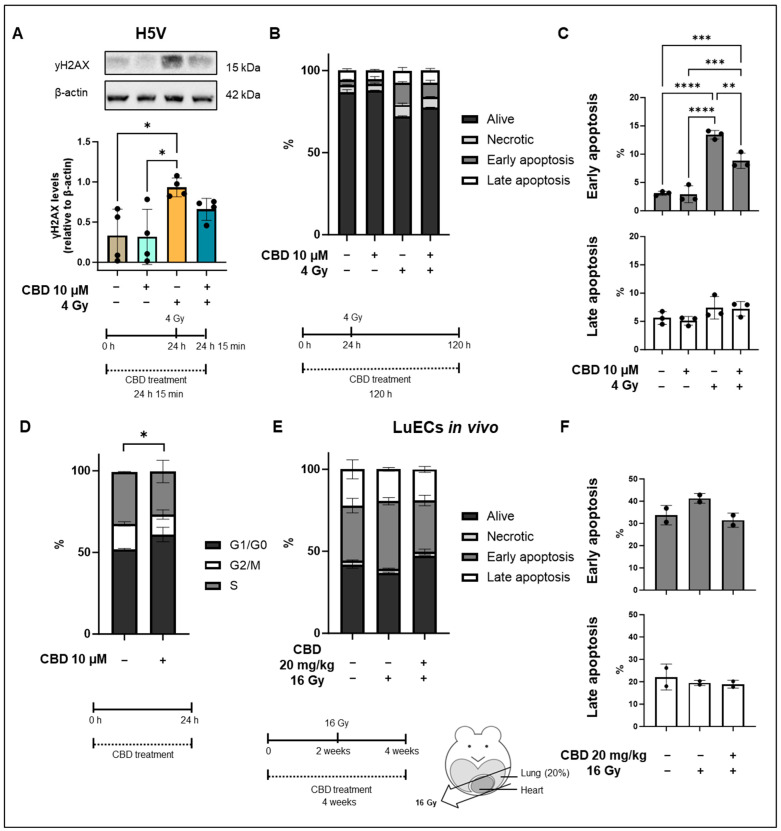
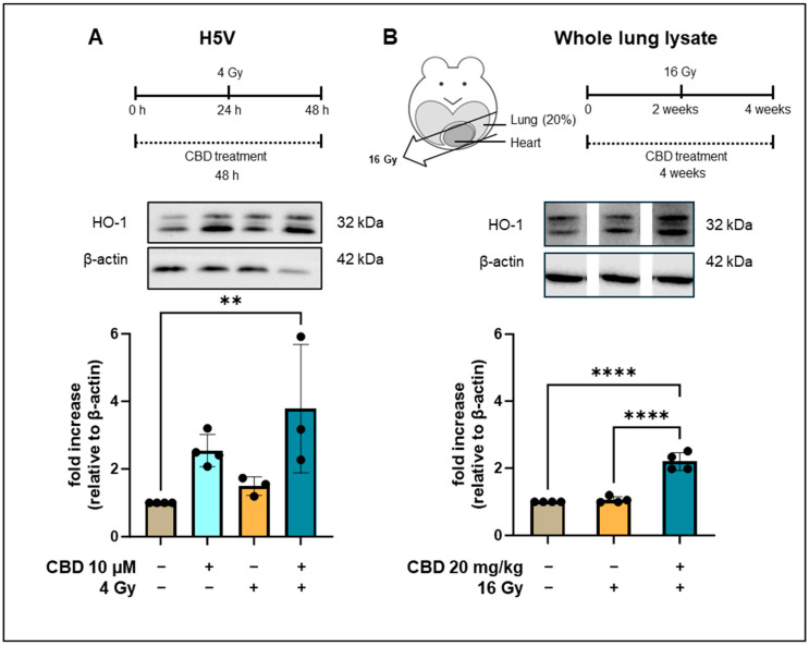
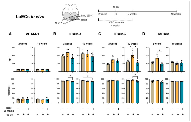
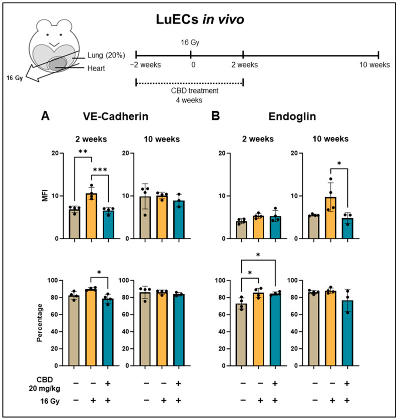
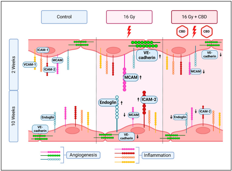
Objective: Radiotherapy, which is commonly used for the local control of thoracic cancers, also induces chronic inflammatory responses in the microvasculature of surrounding normal tissues such as the lung and heart that contribute to fatal radiation-induced lung diseases (RILDs) such as pneumonitis and fibrosis. In this study, we investigated the potential of cannabidiol (CBD) to attenuate the irradiation damage to the vasculature. Methods: We investigated the ability of CBD to protect a murine endothelial cell (EC) line (H5V) and primary lung ECs isolated from C57BL/6 mice from irradiation-induced damage in vitro and lung ECs (luECs) in vivo, by measuring the induction of oxidative stress, DNA damage, apoptosis (in vitro), and induction of inflammatory and pro-angiogenic markers (in vivo). Results: We demonstrated that a non-lethal dose of CBD reduces the irradiation-induced oxidative stress and early apoptosis of lung ECs by upregulating the expression of the cytoprotective mediator heme-oxygenase-1 (HO-1). The radiation-induced increased expression of inflammatory (ICAM-2, MCAM) and pro-angiogenic (VE-cadherin, Endoglin) markers was significantly reduced by a continuous daily treatment of C57BL/6 mice with CBD (i.p. 20 mg/kg body weight), 2 weeks before and 2 weeks after a partial irradiation of the lung (less than 20% of the lung volume) with 16 Gy. Conclusions: CBD has the potential to improve the clinical outcome of radiotherapy by reducing toxic side effects on the microvasculature of the lung.

Keywords: cannabidiol (CBD); inflammation; oxidative stress; radiation-induced lung disease; radiotherapy.


Conflict of interest statement

The authors declare no conflicts of interest.

Figures

Figure A1
Figure A1
(a) Channel slides seeded with lung ECs (luECs in vivo) were perfused with 1 × 10⁶ fluorescently labelled, activated leukocytes under unidirectional flow conditions for 30 min at a flow rate of 1 dyn/cm2. The complete channel is marked with a purple square. (b) View of one analyzed area (marked in red) of a channel (purple) at a magnification of 10×. Adherent leukocytes (green) were determined by automatic particle counting using ImageJ software in all areas of the channel (purple). Attached leukocytes (marked with green circles) were identified by color (green), size (above 5 pixels), and round shape (0.5 to 1.0).
Figure A2
Figure A2
(a) ROS levels in H5V cells 24 h after irradiation: untreated (control, brown column), CBD treatment (10 µM, 48 h, light blue column), irradiation (4 Gy, yellow column), CBD treatment (10 µM, 48 h), and irradiation 24 h after CBD treatment (dark blue column), (n = 5). (b) Clonogenic cell survival of H5V cells 24 h after irradiation: untreated (control), CBD treatment (10 µM, 48 h), irradiation (4 Gy), CBD treatment (10 µM, 48 h), and irradiation 24 h after CBD treatment, (n = 4). The number of colonies in each pre-treatment group was determined on day 7 after seeding; * p < 0.05, ** p < 0.01 and *** p < 0.001; mean fold change ± SD.
Figure A3
Figure A3
Heme oxygenase-1 (HO-1) levels in H5V including NAC treatment. Treatment schedule of HV5 cells treated with CBD/NAC for 48 h and an irradiation with 4 Gy after 24 h measured by western blot. Untreated (lane 1), treated with CBD for 48 h (lane 2), irradiated (4 Gy, 24 h after CBD treatment; lane 3), treated with CBD for 48 h and irradiation (4 Gy, 24 h after CBD treatment; lane 4), treated with NAC for 48 h (lane 5), treated with NAC and CBD for 48 h (lane 6). The fold increase relative to β-actin is from 1 to 4 independent experiments (n = 1–4); statistical analysis was not performed for lanes 5 and 6. Data are mean values ± SD; ** p < 0.01.
Figure A4
Figure A4
Representative gating strategy of CD31+/CD45- lung ECs (luECs in vivo); debris was excluded in SSC-FSC; live cells were gated based on their propidium iodide negativity; endothelial cells (ECs) were selected by their CD31 positivity and CD45 negativity (Q7); markers of interest within EC-population were stained with FITC or PE. The percentage of positively stained cells in CD31+/CD45- lung ECs (luECs in vivo) was determined; MFI was determined by median signal strength of the FITC- or PE-positive EC population; all gates were set based on unstained control samples.
Figure A5
Figure A5
Leukocytes were isolated and cryopreserved from spleens of C57BL/6 mice 2 weeks after partial irradiation of the lungs (20% of lung volume). (a) Lung ECs (luECs in vivo) were isolated 10 weeks after partial irradiation of the lung by a CD31+ selection. Primary lung ECs (luECs in vivo) were cultured in channel slides until confluency was reached and perfused with thawed CFDA-labelled leukocytes (stimulated with IL-2 (100 IU/mL) and Hsp70-peptide TKD (2 µg/mL) for 4–5 days) from the matching treatment group for 30 min. (b) Attached leukocytes were counted based on fluorescent CFDA signal; n = 4; mean numbers of attached leukocytes ± SD.
Figure A6
Figure A6
Expression density (mean fluorescence intensity, MFI) and percentage of lung ECs (luECs in vivo) from C57BL/6 mice cells expressing antigens associated with angiogenesis (Integrin β-3) and repair (CD34) mechanisms. Integrin β-3 (a) and CD34 (b) expression on CD31+/CD45 lung ECs (luECs in vivo) isolated from C57BL/6 mice, 2 and 10 weeks after sham irradiation (0 Gy), partial irradiation of the lung (20% of the total lung volume) with 16 Gy, or partial irradiation of the lung after treatment with CBD (i.p. 20 mg/kg body weight per day for 4 weeks, 2 weeks before and 2 weeks after irradiation); data represent mean values ± SD of 3–4 mice (n = 3-4); * p < 0.05, ** p < 0.01 and *** p < 0.001.
Figure 1
Figure 1
Normalized ROS levels in H5V cells and primary lung ECs (luECs in vitro) upon treatment with CBD, NAC, or both drugs for 24 h followed by an in vitro irradiation (0, 4, 6 Gy). (A) ROS levels in H5V cells determined by DCFDA fluorescence measurements after treatment (24 h) with CBD (10 µM), NAC (100 nM), or both drugs at t0, and 15 min (t1) and 75 min (t2) after sham irradiation (0 Gy). (B) ROS levels in H5V cells after treatment (24 h) with CBD (10 µM), NAC (100 nM), or both drugs 15 min (t1) and 75 min (t2) after irradiation with 4 Gy. (C) ROS levels in H5V cells after treatment (24 h) with CBD (10 µM), NAC (100 nM), or both drugs 15 min (t1) and 75 min (t2) after irradiation with 6 Gy (n = 3–4). (D) ROS levels in primary lung ECs (luECs in vitro) after treatment (24 h) with CBD (10 µM) followed by an in vitro irradiation with 0 Gy (sham) and 4 Gy at t0, t1, and t2; (n = 3); all values are normalized to the respective control values at t0; * p < 0.05, ** p < 0.01, *** p < 0.001 and **** p < 0.0001; data represent mean ROS levels ± SD.
Figure 2
Figure 2
yH2AX levels and apoptosis in H5V cells and primary lung ECs (luECs in vivo) after treatment with CBD and/or irradiation. (A) Representative Western blot of yH2AX and relative yH2AX levels in H5V cells that were untreated (first lane), treated with CBD (10 µM, 24 h; lane 2), treated with irradiation (4 Gy; lane 3), or with CBD (10 µM, 24 h) followed by irradiation (4 Gy; lane 4). All yH2AX levels were determined 15 min after irradiation (4 Gy) in H5V cells. The data represent mean values of 4 independent experiments (n = 4). (B) Percentage of living (alive), necrotic, early (C) and late (C) apoptotic H5V cells determined on day 4 by Annexin/PI staining after no treatment (column 1), CBD treatment (10 µM, 24 h; column 2), irradiation (4 Gy; column 3) and a treatment with CBD (10 µm, 24 h) prior to irradiation (4 Gy; column 4). Data are means ± SD from 3 independent experiments (n = 3); * p < 0.05, ** p < 0.01, *** p < 0.001 and **** p < 0.0001. (D) Cell cycle distribution of H5V cells kept untreated (left column) or 24 h after treatment with 10 µM CBD (right column). Data are means ± SD from 3 independent experiments (n = 3); * p < 0.05; significant difference relates to cells in the G1 phase. (E) Percentage of living (alive), necrotic, early (F) and late (E) apoptotic lung ECs (luECs in vivo) determined 2 weeks after in vivo irradiation by Annexin/PI staining after no treatment (column 1), CBD treatment (20 mg/kg body weight per day, 4 weeks; column 2), CBD-treatment (20 mg/kg body weight per day, 4 weeks) prior to irradiation (16 Gy after 2 weeks CBD treatment; column 3). Data are means from 2 independent experiments (n = 2). Original western blots are presented in Supplementary Materials.
Figure 3
Figure 3
Heme oxygenase-1 (HO-1) levels in H5V and whole lung lysates of C57BL/6 mice. (A) Treatment schedule of HV5 cells treated with CBD for 48 h and an irradiation with 4 Gy after 24 h. Representative Western blot of HO-1 and β-actin levels in H5V cells: untreated (lane 1), treated with CBD for 48 h (lane 2), irradiated (4 Gy, 24 h after CBD treatment; lane 3), treated with CBD for 48 h and irradiation (4 Gy, 24 h after CBD treatment; lane 4). The fold increase relative to β-actin is from 3 to 4 independent experiments (n = 3–4). (B) Treatment schedule of C57BL/6 mice receiving either CBD for 4 weeks (20 mg/kg body weight per day) or a partial lung irradiation (16 Gy) after 2 weeks. Representative Western blots of HO-1 and β-actin levels (images spliced from same blot) in whole lung lysates of C57BL/6 mice kept untreated (lane 1), after partial lung irradiation (16 Gy, lane 2), or after treatment with CBD (20 mg/kg body weight per day) for 4 weeks and irradiation (partial lung irradiation 16 Gy after 2 weeks, lane 3). Data are mean values ± SD of 4 C57BL/6 mice; ** p < 0.01 and **** p < 0.0001. Original western blots are presented in Supplementary Materials.
Figure 4
Figure 4
Expression density (mean fluorescence intensity, MFI) and percentage of cells positively stained for inflammatory markers on lung ECs (luECs in vivo) from C57BL/6 mice. (A) VCAM-1, (B) ICAM-1, (C) ICAM-2, and (D) and MCAM expression on CD31+/CD45- ECs from the lungs of C57BL/6 mice, 2 and 10 weeks after sham irradiation (0 Gy, brown column), partial irradiation of the lung (20% of the total lung volume) with 16 Gy (yellow column), or partial lung irradiation after a treatment with CBD (i.p. 20 mg/kg body weight per day for 4 weeks, 2 weeks before and 2 weeks after lung irradiation, blue column); data show mean values ± SD of 3–4 mice (n = 3–4); * p < 0.05.
Figure 5
Figure 5
Expression density (mean fluorescence intensity, MFI) and percentage of lung ECs (luECs in vivo) from C57BL/6 mice cells expressing antigens associated with repair and angiogenesis mechanisms. (A) VE-cadherin and (B) Endoglin expression on CD31+/CD45 lung ECs (luECs in vivo) isolated from C57BL/6 mice, 2 and 10 weeks after sham irradiation (0 Gy, brown column), partial irradiation of the lung (20% of the total lung volume) with 16 Gy (yellow column), or partial lung irradiation after a treatment with CBD (i.p. 20 mg/kg body weight per day for 4 weeks, 2 weeks before and 2 weeks after lung irradiation, blue column); data represent mean values ± SD of 3–4 mice (n = 3–4); * p < 0.05, ** p < 0.01 and *** p < 0.001.
Figure 6
Figure 6
Changes in the expression of inflammatory (VCAM-1, ICAM-1, ICAM-2, MCAM) and angiogenic (VE-cadherin, Endoglin, MCAM) markers on primary lung ECs (luECs in vivo) 2 and 10 weeks after partial irradiation of the lung with 16 Gy, and the recovery effect of a 4-week treatment with CBD, beginning 2 weeks prior to irradiation. Created in BioRender [34].

References

    1. Baskar R., Dai J., Wenlong N., Yeo R., Yeoh K.W. Biological response of cancer cells to radiation treatment. Front. Mol. Biosci. 2014;1:24. doi: 10.3389/fmolb.2014.00024. - DOI - PMC - PubMed
    1. Rakotomalala A., Escande A., Furlan A., Meignan S., Lartigau E. Hypoxia in Solid Tumors: How Low Oxygenation Impacts the “Six Rs” of Radiotherapy. Front. Endocrinol. 2021;12:742215. doi: 10.3389/fendo.2021.742215. - DOI - PMC - PubMed
    1. Giuranno L., Ient J., De Ruysscher D., Vooijs M.A. Radiation-Induced Lung Injury (RILI) Front. Oncol. 2019;9:877. doi: 10.3389/fonc.2019.00877. - DOI - PMC - PubMed
    1. Beach T.A., Groves A.M., Williams J.P., Finkelstein J.N. Modeling radiation-induced lung injury: Lessons learned from whole thorax irradiation. Int. J. Radiat. Biol. 2020;96:129–144. doi: 10.1080/09553002.2018.1532619. - DOI - PMC - PubMed
    1. Hamada N., Kawano K.I., Yusoff F.M., Furukawa K., Nakashima A., Maeda M., Yasuda H., Maruhashi T., Higashi Y. Ionizing Irradiation Induces Vascular Damage in the Aorta of Wild-Type Mice. Cancers. 2020;12:3030. doi: 10.3390/cancers12103030. - DOI - PMC - PubMed

LinkOut - more resources