. 2024 Nov 3;16(21):3711.
doi: 10.3390/cancers16213711.

Enhancing Anti-PD-1 Immunotherapy by Targeting MDSCs via Hepatic Arterial Infusion in Breast Cancer Liver Metastases

Affiliations

Enhancing Anti-PD-1 Immunotherapy by Targeting MDSCs via Hepatic Arterial Infusion in Breast Cancer Liver Metastases

Minhyung Kim et al. Cancers (Basel). .

Abstract

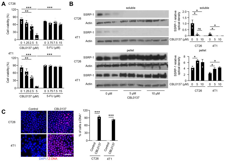
Background: Surgery, chemotherapy, and radiation often have limited utility for advanced metastatic disease in the liver, and despite its promising activity in select cancers, PD-1 blockade therapy similarly has minimal benefit in this setting. Curaxin, CBL0137, is an experimental anti-cancer drug that disrupts the binding of DNA to histones, destabilizes chromatin, and induces Z-DNA formation which may stimulate anti-tumor immune responses.

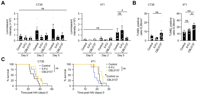
Methods: Murine cell lines of colon (CT26) and breast (4T1) cancer were interrogated for survival and CBL0137-associated DNA changes in vitro. Immunocompetent models of liver metastases followed by CBL0137 hepatic arterial infusion (HAI) were used to examine in vivo tumor cell DNA alterations, treatment responses, and the immune contexture associated with CBL0137, both alone and in combination with anti-PD-1 therapy.

Results: CBL0137 induced immediate changes to favor tumor cell death in vitro and in vivo with an efficient tumor uptake via the HAI route. Toxicity to CBL0137 was minimal and anti-tumor treatment effects were more efficient with HAI compared to intravenous delivery. Immune effects were pronounced with CBL0137 HAI with concurrent depletion of a specific population of myeloid-derived suppressor cells and maintenance of effector T cell populations.

Conclusions: Combination of CBL0137 HAI with PD-1 blockade improved survival in 4T1 tumors but not in CT26 tumors, and therapeutic efficacy relies on the finding of simultaneous and targeted depletion of myeloid-derived suppressor cells and skewing of T cell populations to produce synergy with PD-1 blockade therapy.

Keywords: Z-DNA; hepatic arterial infusion (HAI); immunotherapy; liver tumor; myeloid-derived suppressor cells (MDSCs).


Conflict of interest statement

The authors report no competing interests.

Figures

Figure 1
Figure 1
A schematic diagram illustrating the mechanism of action involving chromatin decondensation and the chemical structure of CBL0137. Z-DNA formation occurs following the interaction of CBL0137 with chromatin, leading to histone disassembly.
Figure 2
Figure 2
A schematic illustration and representative photographs of HAI via the superior pancreaticoduodenal artery (SPDA). (A) A diagram of the HAI procedure via the SPDA. (B) A representative photograph of the vascular anatomy before cannulation. (C) A representative photograph after catheter placement (*) in the SPDA.
Figure 3
Figure 3
CBL0137 rapidly limits proliferation of CT26 and 4T1 tumor cells in vitro by altering DNA/histone interactions. (A) In vitro cell viability was significantly reduced by a 15 min treatment with CBL0137 as compared to 5FU (n = 5). Representative data among 3 independent experiments. **: p < 0.01; ***: p < 0.001 from Student’s t-test; error bar: standard error of the mean. (B) Western blot analysis of SSRP 1 redistribution after in vitro treatment with different doses of CVB0137. As a marker for CBL0137 activity, SSRP1 loss in the soluble fraction of nuclear proteins and accumulation in the pelleted fraction of proteins from tumor cells was noted in vitro. Representative data between 2 independent experiments. Results were quantified using 1.53v ImageJ. n = 3; *: p < 0.05 from Student’s t-test; error bar: standard error of the mean, ns: not significant. (C) Immunofluorescence (IF) of Z-DNA in cancer cells. CT26 and 4T1 cells were treated with either the control (D5W) or CBL0137 for 15 min in vitro and then were stained with Z-DNA antibodies showing marked induction of Z-DNA formation. Representative IF figures among 3 independent experiments. n = 3; bar is 50 µm; ***: p < 0.001 from Student’s t-test; error bar: standard error of the mean.
Figure 4
Figure 4
HAI with CBL0137 induces distinct chromatin decondensation with Z-DNA formation in vivo. (A) Western blot analysis of SSRP 1 redistribution after in vivo treatment via CBL0137 HAI. HAI with CBL0137 induced DNA/histone changes as noted by loss of SSRP-1 in soluble fractions and appearance in the pellets of 4T1 and CT26 liver tumors. Single experiment. (B) Z-DNA detection using IF after in vivo HAI. In vivo HAI with CBL0137 induced nuclear changes as depicted by the generation of Z-DNA as compared to control (D5W) or 5-FU. Representative IF figures among 3 independent experiments. n = 6; ***: p < 0.001 from Student’s t-test; error bar: standard error of the mean. (C) Suppression of NF-κB expression 4 h after CBL0137 HAI compared to the control HAI group. Representative data from 2 independent experiments. n = 3, *; p < 0.05, ***; p < 0.001 from Student’s t-test, error bar; standard error of the mean.
Figure 5
Figure 5
CBL0137 HAI produces different antitumor responses in CT26 and 4T1 liver metastases. (A) Luminescence 5 days after HAI with CBL0137 was reduced significantly in 4T1 liver tumors, but not CT26 liver tumors; representative data among 3 independent experiments, CT26 (n = 6) and 4T1 (n = 5). (B) Numbers of TUNEL-positive cells in tumor nodules. A trend to greater numbers of apoptotic cells and a statistical increase in apoptotic cells are identified by TUNEL stain 5 days after HAI treatment with CBL0137 in CT26 and 4T1 liver metastases. Pooled data with 3 independent experiments with CT26 tumor (n = 10) and 4T1 (n = 8) tumor. Each value represented an average of 3 slides per mouse. *: p < 0.05; **: p < 0.01 from Student’s t-test; error bar: standard error of the mean; ns: not significant. (C) Survival curves after HAI with different treatments. No survival benefit was detected in mice bearing CT26 liver metastasis. Alternatively, survival data showed statistically significant but modest survival benefits in mice bearing 4T1 liver metastasis. Pooled data from 3 independent experiments. *: p < 0.05; ns: not significant; n = 9, using log-rank tests.
Figure 6
Figure 6
CBL0137 HAI exerts favorable changes in immune cell composition of metastatic tumors. (A) Representative IF images of CD8- and Gr-1-positive cells. CBL0137 reduced intratumoral MDSCs in both CT26 and 4T1 tumors. Gr-1+ (red) and CD8+ (green) antibodies 5 days after HAI with control (D5W), 5FU, or CBL0137. The scale bar is 50 μm; the white dashed line denotes the tumor boundary. (B) Numbers of MDSCs in tumor modules after HAI with different treatments in CT26 and 4T1 liver metastatic models. CBL0137 HAI decreased the number of MDSCs in 4T1 liver tumors but had no impact on the already low numbers of MDSCs in CT26 liver tumors. ***: p < 0.001 from Student’s t-test; error bar: standard error of the mean; ns: not significant. (C) Numbers of CD8+ T cells in tumor nodules after HAI. CD8+ cells tended to increase in CT26 tumor nodules and significantly increased in 4T1 tumor nodules from CBL0137 HAI. #; number, *: p < 0.05; **: p < 0.01; ***: p < 0.001 from Student’s t-test; error bar: standard error of the mean; ns: not significant. (D) The ratios of MDSCs and CD8+ T cells in tumor nodules. CBL0137 induces favorable statistically significant MDSC/CD8+ T cell ratios in CT26 and 4T1 tumors 5 days after HAI. Representative photos and pooled data from 3 independent experiments, with each value representing an average of 3 slides per mouse. *: p < 0.05; **: p < 0.01; ***: p < 0.001; n = 10, from Student’s t-test; error bar: standard error of the mean.
Figure 7
Figure 7
PD-1 blockade therapy augments survival of CBL0137 HAI. (A) Survival curves with anti-PD-1 therapy after HAI. Overall survival was improved, and cures generated by the addition of anti-PD-1 in HAI CBL0137 and 5-FU groups in CT26 tumor-bearing mice. And overall survival was significantly extended by the addition of anti-PD-1 therapy to CBL0137 HAI in 4T1 tumor-bearing mice compared to PD-1 alone. Survival data were acquired after each treatment with mice bearing 4T1 and CT26 liver metastases. Pooled data from 2 different experiments. **: p < 0.01; ns: not significant; n ≥ 5, using log-rank tests. (B) Compositions of CD4+/CD8+ naïve, central memory, and effector memory T cell populations. T cell-activated statuses were analyzed as naïve (CD62L+CD44), central memory (CM; CD62L+CD44+), and effector memory (EM; CD62LCD44+) cell populations 5 days after each treatment and showed significant skewing toward effector memory subsets in combined CBL0137 and anti-PD-1 treatment in 4T1 mice. Pooled data from 2 different experiments. *: p < 0.05; ***: p < 0.001; ns: not significant; n ≥ 4, from Student’s t-test.
Figure 8
Figure 8
CBL0137 HAI selectively depletes Il1b and CD84-expressing hepatic MDSC populations. (A) ScRNAseq was performed with leukocytes from the liver 5 days after HAI with either vehicle or CBL0137. Cell clusters were characterized after vehicle or CBL0137 treatment. (B) Subcategorized neutrophil and monocyte populations based on ImmGen data. (C) Neutrophil and monocyte populations were identified with Il1b and CD84 genes after vehicle or CBL137 HAI. (D) Violin plots showing different levels of Il1b and Cd84 gene expression in existing MDSC populations after vehicle or CBL0137 treatment. (E) Western blot for the Z-DNA-sensing protein ZBP1 and a downstream molecule (RIPK3) in MDSCs recovered under different conditions (as depicted) shows consistently high levels of ZBP1 and RIPK3 in all MDSCs recovered. (F) IF images of Z-DNA in T cells and MDSCs. After 1 h of exposure to CBL0137 in vitro, Z-DNA is extensively detected in MDSCs but not T cells. Representative IF figures among 2 independent experiments. n = 3; **: p < 0.01 from Student’s t-test; error bar: standard error of the mean.
Figure 8
Figure 8
CBL0137 HAI selectively depletes Il1b and CD84-expressing hepatic MDSC populations. (A) ScRNAseq was performed with leukocytes from the liver 5 days after HAI with either vehicle or CBL0137. Cell clusters were characterized after vehicle or CBL0137 treatment. (B) Subcategorized neutrophil and monocyte populations based on ImmGen data. (C) Neutrophil and monocyte populations were identified with Il1b and CD84 genes after vehicle or CBL137 HAI. (D) Violin plots showing different levels of Il1b and Cd84 gene expression in existing MDSC populations after vehicle or CBL0137 treatment. (E) Western blot for the Z-DNA-sensing protein ZBP1 and a downstream molecule (RIPK3) in MDSCs recovered under different conditions (as depicted) shows consistently high levels of ZBP1 and RIPK3 in all MDSCs recovered. (F) IF images of Z-DNA in T cells and MDSCs. After 1 h of exposure to CBL0137 in vitro, Z-DNA is extensively detected in MDSCs but not T cells. Representative IF figures among 2 independent experiments. n = 3; **: p < 0.01 from Student’s t-test; error bar: standard error of the mean.
Figure 9
Figure 9
ScRNAseq data reveal differences in cell clusters and gene expressions. Isolated leukocytes from the liver 5 days after HAI with either vehicle or CBL0137 were analyzed to (A) characterize cell populations and (B) different gene expressions in MDSCs, neutrophils, monocytes, and T and NK cells.

Similar articles

References

    1. Abdalla E.K., Vauthey J.N., Ellis L.M., Ellis V., Pollock R., Broglio K.R., Hess K., Curley S.A. Recurrence and outcomes following hepatic resection, radiofrequency ablation, and combined resection/ablation for colorectal liver metastases. Ann. Surg. 2004;239:818–825. doi: 10.1097/01.sla.0000128305.90650.71. discussion 825–817. - DOI - PMC - PubMed
    1. Power D.G., Healey-Bird B.R., Kemeny N.E. Regional chemotherapy for liver-limited metastatic colorectal cancer. Clin. Color. Cancer. 2008;7:247–259. doi: 10.3816/CCC.2008.n.032. - DOI - PubMed
    1. DeWitt J., Yu M., Al-Haddad M.A., Sherman S., McHenry L., Leblanc J.K. Survival in patients with pancreatic cancer after the diagnosis of malignant ascites or liver metastases by EUS-FNA. Gastrointest. Endosc. 2010;71:260–265. doi: 10.1016/j.gie.2009.08.025. - DOI - PubMed
    1. Jerraya H., Saidani A., Khalfallah M., Bouasker I., Nouira R., Dziri C. Management of liver metastases from gastric carcinoma: Where is the evidence? Tunis Med. 2013;91:1–5. - PubMed
    1. Ursaru M., Jari I., Negru D., Scripcariu V. Local and distant recurrences—A comparative study on conservative and radical surgery for breast cancer. Chirurgia. 2015;110:38–42. - PubMed

Grants and funding

LinkOut - more resources