. 2024 Nov 4;16(21):3100.
doi: 10.3390/polym16213100.

Polypiperazine-Based Micelles of Mixed Composition for Gene Delivery

Affiliations

Polypiperazine-Based Micelles of Mixed Composition for Gene Delivery

Rumena Stancheva et al. Polymers (Basel). .

Abstract

We introduce a novel concept in nucleic acid delivery based on the use of mixed polymeric micelles (MPMs) as platforms for the preparation of micelleplexes with DNA. MPMs were prepared by the co-assembly of a cationic copolymer, poly(1-(4-methylpiperazin-1-yl)-propenone)-b-poly(d,l-lactide), and nonionic poly(ethylene oxide)-b-poly(propylene oxide)-b-poly(ethylene oxide) block copolymers. We hypothesize that by introducing nonionic entities incorporated into the mixed co-assembled structures, the mode and strength of DNA binding and DNA accessibility and release could be modulated. The systems were characterized in terms of size, surface potential, buffering capacity, and binding ability to investigate the influence of composition, in particular, the poly(ethylene oxide) chain length on the properties and structure of the MPMs. Endo-lysosomal conditions were simulated to follow the changes in fundamental parameters and behavior of the micelleplexes. The results were interpreted as reflecting the specific structure and composition of the corona and localization of DNA in the corona, predetermined by the poly(ethylene oxide) chain length. A favorable effect of the introduction of the nonionic block copolymer component in the MPMs and micelleplexes thereof was the enhancement of biocompatibility. The slight reduction of the transfection efficiency of the MPM-based micelleplexes compared to that of the single-component polymer micelles was attributed to the premature release of DNA from the MPM-based micelleplexes in the endo-lysosomal compartments.

Keywords: PEO-PPO-PEO copolymers; cationic copolymers; gene/DNA delivery; internalization/transfection; micelleplexes; mixed polymeric micelles; polyplexes.


Conflict of interest statement

The authors declare no conflicts of interest.

Figures

Figure 1
Figure 1
Chemical structures of (a) polypiperazine homopolymers according to ref. [34] (R = CH3, C2H5, C3H7, C4H9); (b) PMPP-PLA and (c) PEO-PPO-PEO block copolymers. n and m values represent the degree of polymerization of hydrophilic and hydrophobic blocks of the copolymers and are given in Table S1.
Figure 2
Figure 2
Schematic illustration of the structure of MPMs based on PMPP-PLA and PEO-PPO-PEO block copolymers.
Figure 3
Figure 3
Buffering capacity of SCPMs and MPMs based on PMPP-PLA and PEO-PPO-PEO block copolymers given as (a) pH vs. volume of 0.1 M HCl curves and (b) reciprocal value of the slope of curves from (a).
Figure 4
Figure 4
Ethidium bromide fluorescence quenching in micelleplexes formed from SCPMs and MPMs based on PMPP-PLA and PEO-PPO-PEO block copolymers followed at pH 7 (a) and after incubation for 24 h in endo–lysosomal conditions (pH 4, 37 °C) (b). The fluorescence intensity of ethidium bromide in pure DNA was taken as 100%.
Figure 5
Figure 5
Variations of hydrodynamic radius, Rh, with the N/P ratio of micelleplexes formed from PMPP-PLA SCPMs (a) and PMPP-PLA/L64 (b), PMPP-PLA/P65 (c), and PMPP-PLA/F77 (d) MPMs. Measurements were made at pH 7 (closed symbols) and after incubation for 24 h in endo–lysosomal conditions (pH 4, 37 °C, open symbols).
Figure 5
Figure 5
Variations of hydrodynamic radius, Rh, with the N/P ratio of micelleplexes formed from PMPP-PLA SCPMs (a) and PMPP-PLA/L64 (b), PMPP-PLA/P65 (c), and PMPP-PLA/F77 (d) MPMs. Measurements were made at pH 7 (closed symbols) and after incubation for 24 h in endo–lysosomal conditions (pH 4, 37 °C, open symbols).
Figure 6
Figure 6
Variations of ζ potential with the N/P ratio of micellplexes formed from PMPP-PLA SCPMs (a) and PMPP-PLA/L64 (b), PMPP-PLA/P65 (c), and PMPP-PLA/F77 (d) MPMs. Measurements were made at pH 7 (closed symbols) and after incubation for 24 h in endo–lysosomal conditions (pH 4, 37 °C, open symbols).
Figure 6
Figure 6
Variations of ζ potential with the N/P ratio of micellplexes formed from PMPP-PLA SCPMs (a) and PMPP-PLA/L64 (b), PMPP-PLA/P65 (c), and PMPP-PLA/F77 (d) MPMs. Measurements were made at pH 7 (closed symbols) and after incubation for 24 h in endo–lysosomal conditions (pH 4, 37 °C, open symbols).
Figure 7
Figure 7
Schematic illustration of the structure of micelleplexes prepared from SCPMs and MPMs based on PMPP-PLA and PEO-PPO-PEO block copolymers.
Figure 8
Figure 8
Determination of the cytotoxic effect of the tested micelles (a) and their respective micelleplexes (b) on L929 cells was conducted by dose–response curves using the “concentration vs. normalized response (variable slope)” algorithm in GraphPad Prism v.8 software. Cell viability is indicated as a percentage relative to untreated control cells. The presented data include the mean ± SD of four replicates from two separate experiments. The red line corresponds to 70% cell viability.
Figure 9
Figure 9
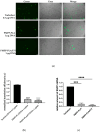
Internalization capacity of tested micelleplexes. (a) Representative microscopic images showing H1299 cells treated with the examined micelleplexes at pH = 7, with a final salmon sperm DNA concentration of 1 μg mL−1 for 24 h. The scale bar corresponds to 40 μm. (b) Quantitative analysis of EtBr fluorescence intensity was conducted using the Fiji image processing package. At least four images for each tested micelleplex from two independent experiments were assessed. The values represent the means ± SD. Statistical analysis was performed using a one-way ANOVA Tukey’s multiple comparisons test to compare the mean of each column with the mean of every other column. No statistically significant differences were observed.
Figure 10
Figure 10
Transfection capacity of tested micelleplexes. (a) Representative microscopic images showing H1299 cells transfected with 0.5 μg pEGFP-N1 using Turbofect (positive control) or treated with micelleplexes containing pEGFP-N1 at a final DNA concentration of 1 μg mL−1 for 6 h. At least three images for each condition from two independent experiments were assessed. The scale bar corresponds to 40 μm. (b) Bar graphs present transfected cells vs. total number of cells from each condition. (c) Relative amounts of EGFP gene mRNA after 24 h incubation in H1299 cells transfected with pEGFP-N1 using Turbofect (positive control) or treated with micelleplexes containing the same plasmid DNA. The relative amounts of EGFP gene mRNA in micelleplexes are normalized to the relative amount of EGFP gene mRNA in the positive control (Turbofect). Statistical analysis was performed using a one-way ANOVA Dunnett’s multiple comparisons test to compare the mean of each column with the mean of control column (Turbofect). Probability values indicating significance were considered at the *** p < 0.005, **** p < 0.001.

References

    1. Witten J., Hu Y., Langer R., Anderson D.G. Recent advances in nanoparticulate RNA delivery systems. Proc. Natl. Acad. Sci. USA. 2024;121:e2307798120. doi: 10.1073/pnas.2307798120. - DOI - PMC - PubMed
    1. Ratner M. First gene therapy approved. Nat. Biotechnol. 2012;30:1153
    1. Ma C.C., Wang Z.L., Xu T., He Z.Y., Wei Y.Q. The approved gene therapy drugs worldwide: From 1998 to 2019. Biotechnol. Adv. 2020;40:107502. - PubMed
    1. Nayerossadat N., Maedeh T., Ali P.A. Viral and nonviral delivery systems for gene delivery. Adv. Biomed. Res. 2012;1:27. doi: 10.4103/2277-9175.98152. - DOI - PMC - PubMed
    1. Lentz T.B., Gray S.J., Samulski R.J. Viral vectors for gene delivery to the central nervous system. Neurobiol. Dis. 2012;48:179. doi: 10.1016/j.nbd.2011.09.014. - DOI - PMC - PubMed

LinkOut - more resources