. 2024 Oct 26;14(21):2389.
doi: 10.3390/diagnostics14212389.

Sarcoid Nodule or Lung Cancer? A High-Resolution Computed Tomography-Based Retrospective Study of Pulmonary Nodules in Patients with Sarcoidosis

Affiliations

Sarcoid Nodule or Lung Cancer? A High-Resolution Computed Tomography-Based Retrospective Study of Pulmonary Nodules in Patients with Sarcoidosis

Chiara Catelli et al. Diagnostics (Basel). .

Abstract

Background: The objective of this retrospective study was to compare the characteristics of sarcoid nodules and neoplastic nodules using high-resolution computed tomography (HRCT) in sarcoidosis patients. Methods: This is a single-center retrospective study. From 2010 to 2023, among 685 patients affected by pulmonary sarcoidosis, 23 patients developed pulmonary nodules of a suspicious malignant nature. The HRCT characteristics of biopsy-proven malignant (Group A) vs. inflammatory (Group B) nodules were analyzed and compared. Results: A significant difference was observed between the groups in terms of age (p = 0.012). With regard to HRCT features, statistical distinctions were observed in the appearance of the nodule, more frequently spiculated in the case of lung cancer (p < 0.01), in the diameter of the nodule (Group A: 23.5 mm; Group B: 12.18 mm, p < 0.02), in the median nodule density (Group A: 60.0 HU, Group B: -126.7 HU, p < 0.01), and in the number of pulmonary nodules, as a single parenchymal nodule was more frequently observed in the neoplastic patient group (p = 0.043). In Group A, the 18-PET-CT demonstrated hilar/mediastinal lymphadenopathy in 100% of cases; histology following surgery did not report any cases of malignant lymph node involvement. Conclusions: An accurate clinical evaluation and HRCT investigation are crucial for diagnosing lung cancer in patients with sarcoidosis in order to determine who requires surgical resection. The spiculated morphology of the nodule, greater size, the number of pulmonary nodules, and density using HRCT appear to correlate with the malignant nature of the lesion.

Keywords: HRCT; computed tomography; lung adenocarcinoma; lung cancer; lung nodule; lung resection; lymph nodes; pulmonary sarcoidosis.


Conflict of interest statement

The authors declare no conflicts of interest.

Figures
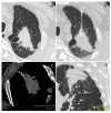
Figure 1
Figure 1
(ad). This is the case of an 80-year-old patient with acinar (40%) and solid (20%) adenocarcinoma with in situ areas of lepidic morphology (40%) and adenocarcinomatous infiltration of the visceral pleura (a), confirmed on biopsy examination (b). The neoplastic lesion has a high density (61.56 HU, (c)) and a size of about 60 mm (d).
Figure 2
Figure 2
(ae). The use of an examination technique with bone plus kernel reconstruction (for the study of lung parenchyma) (a), high spatial resolution (for the study of morphology and margins) (b), and high contrast resolution (for the study of intranodular densities) (c) allow for an accurate characterization of nodular lesions. This type of study makes it possible to obtain images of high diagnostic quality also in coronal (d) and sagittal reconstructions (e), enabling the differential diagnosis of pulmonary nodules.
Figure 3
Figure 3
Decision algorithm on surgical indication in patients with sarcoidosis and suspicious lung nodule (defined as new-onset nodules that persist or grow during radiological follow-up regardless medical therapy) in the absence of a diagnosis.

References

    1. Kelleher D.W., Yaggi M., Homer R., Herzog E.L., Ryu C. A rare presentation of pulmonary sarcoidosis as a solitary lung mass: A case report. J. Med. Case Rep. 2018;12:94. doi: 10.1186/s13256-018-1632-0. - DOI - PMC - PubMed
    1. Bianchi F., Piccioli C., Rosi E., Carobene L., Spina D., Mazzei M., Bartolucci M., Moroni C., Novelli L., Rottoli P., et al. Combined sarcoidosis and idiopathic pulmonary fibrosis (CSIPF): A novel disease phenotype? Respir. Med. 2019;160:105650. doi: 10.1016/j.rmed.2019.02.022. - DOI - PubMed
    1. Margaritopoulos G.A., Proklou A., Lagoudaki E., Voloudaki A., Siafakas N.M., Antoniou K.M. Sarcoidosis in a 65-year-old woman presenting with a lung mass and pericardial effusion: A case report. J. Med. Case Rep. 2012;6:259. doi: 10.1186/1752-1947-6-259. - DOI - PMC - PubMed
    1. Gotway M.B., Tchao N.K., Leung J.W., Hanks D.K., Thomas A.N. Sarcoidosis presenting as an enlarging solitary pulmonary nodule. J. Thorac. Imaging. 2001;16:117–122. doi: 10.1097/00005382-200104000-00010. - DOI - PubMed
    1. Malaisamy S., Dalal B., Bimenyuy C., Soubani A.O. The clinical and radiologic features of nodular pulmonary sarcoidosis. Lung. 2009;187:9–15. doi: 10.1007/s00408-008-9118-2. - DOI - PubMed

LinkOut - more resources