Review
. 2024 Oct 28;14(21):2399.
doi: 10.3390/diagnostics14212399.

Chemotherapy Induced Corneal Changes Assessed by Corneal Confocal Microscopy: A Review

Affiliations
Review

Chemotherapy Induced Corneal Changes Assessed by Corneal Confocal Microscopy: A Review

Eleonora Cosmo et al. Diagnostics (Basel). .

Abstract

The eye, and the cornea in particular, is a common site of chemotherapy induced toxicity, and ocular side effects of both traditional and novel agents have been reported. Corneal confocal microscopy (CCM) is an in vivo technique that allows for the study of all the corneal layers in an easy, non-invasive and reproducible way via the direct visualization of corneal cell morphologies as well as of sub-basal nerve plexus. Thus, it represents a useful way to identify and monitor chemotherapy induced corneal alterations. This work aims to review the use of CCM in identifying corneal toxicity secondary to chemotherapy treatment, as regards both corneal nerves alterations in the setting of chemotherapy induced peripheral neuropathy (CIPN) and other corneal structure changes, particularly involving the corneal epithelium.

Keywords: chemotherapy; cornea; corneal confocal microscopy; corneal epithelium; corneal toxicity; peripheral neuropathy.


Conflict of interest statement

The authors declare no conflicts of interest.

Figures
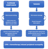
Figure 1
Figure 1
Schematic summary explaining the pathogenic mechanisms of platinum compounds and taxanes induced peripheral neuropathy, which manifests itself even in the corneal sub-basal nerve plexus, thus being visible by means of corneal confocal microscopy.
Figure 2
Figure 2
Images (400 × 400 microns) of the sub-basal nerve plexus of an healthy patient (A) and of a subject undergoing therapy with oxaliplatin (B), captured by means of corneal confocal microscopy (Heidelberg Retina Tomograph III Rostock Corneal Module, Heidelberg, Germany). The plexus of the patient in treatment with oxaliplatin is characterized by a reduced number of thinner nerve fibers compared to that of the healthy subject. Furthermore, there is evidence of immature dendritic cells among the nerve fibers of the patient undergoing chemotherapy treatment.
Figure 3
Figure 3
Schematic summary explaining the on-target and off-target mechanisms exerted by different chemotherapy drugs consisting in antibodies with or without conjugated citotoxin, causing corneal side effects. Epidermal growth factor receptor (EGFR) inhibitors (such as vandetanib, erlotinib, cetuximab and panitumumab) cause direct damage to epithelial cells by inhibiting EGFR with consequent impairment in the normal epithelial turnover and cell migration. Antibody-drug-conjugated (ADC), such as trastuzumab emtansine and depatuxizumab mafodotin, lead to corneal injury mainly via on-target mechanisms, since the antigens target of these ADC (HER-2 for trastuzumab and EGFR for depatuxizumab) are expressed in the corneal epithelium. Whereas, belantamab mafodotin causes corneal changes primarily with off-target mechanisms, since belantamab target (i.e., B-cell maturation antigen) is not expressed in the cornea.
Figure 4
Figure 4
Corneal confocal microscopy images (400 × 400 microns) captured with Heidelberg Retina Tomograph III Rostock Corneal Module (Heidelberg, Germany) of patients undergoing therapy with belantamab mafodotin (belamaf), showing corneal microcyst-like epithelial changes induced by the therapy. Specifically, image in (A) shows hyperreflective opacities within the corneal basal epithelium; image in (B) demonstrates the presence of intraepithelial round microcystic structures consisting of a hyperreflective wall; image in (C) shows a cluster of epithelial hyperreflective deposits, which are also evident in the sub-basal nerve plexus layer (as evident in image in (D)), characterized by reduced and thinner fibers.

References

    1. Sun T., Niu X., He Q., Liu M., Qiao S., Qi R.Q. Development, Efficacy and Side Effects of Antibody–Drug Conjugates for Cancer Therapy (Review) Mol. Clin. Oncol. 2023;18:47. doi: 10.3892/mco.2023.2643. - DOI - PMC - PubMed
    1. Schirrmacher V. From Chemotherapy to Biological Therapy: A Review of Novel Concepts to Reduce the Side Effects of Systemic Cancer Treatment. Int. J. Oncol. 2019;54:407–419. doi: 10.3892/ijo.2018.4661. - DOI - PMC - PubMed
    1. Kunkler A.L., Binkley E.M., Mantopoulos D., Hendershot A.J., Ohr M.P., Kendra K.L., Davidorf F.H., Cebulla C.M. Known and Novel Ocular Toxicities of Biologics, Targeted Agents, and Traditional Chemotherapeutics. Graefe’s Arch. Clin. Exp. Ophthalmol. 2019;257:1771–1781. doi: 10.1007/s00417-019-04337-8. - DOI - PubMed
    1. DelMonte D.W., Kim T. Anatomy and Physiology of the Cornea. J. Cataract Refract. Surg. 2011;37:588–598. doi: 10.1016/j.jcrs.2010.12.037. - DOI - PubMed
    1. Oliveira-Soto L., Efron N. Morphology of Corneal Nerves Using Confocal Microscopy. Cornea. 2001;20:374–384. doi: 10.1097/00003226-200105000-00008. - DOI - PubMed

LinkOut - more resources