

Digital Holographic Microscopy in Veterinary Medicine-A Feasibility Study to Analyze Label-Free Leukocytes in Blood and Milk of Dairy Cows
- PMID: 39518879
- PMCID: PMC11544890
- DOI: 10.3390/ani14213156
Digital Holographic Microscopy in Veterinary Medicine-A Feasibility Study to Analyze Label-Free Leukocytes in Blood and Milk of Dairy Cows
Abstract
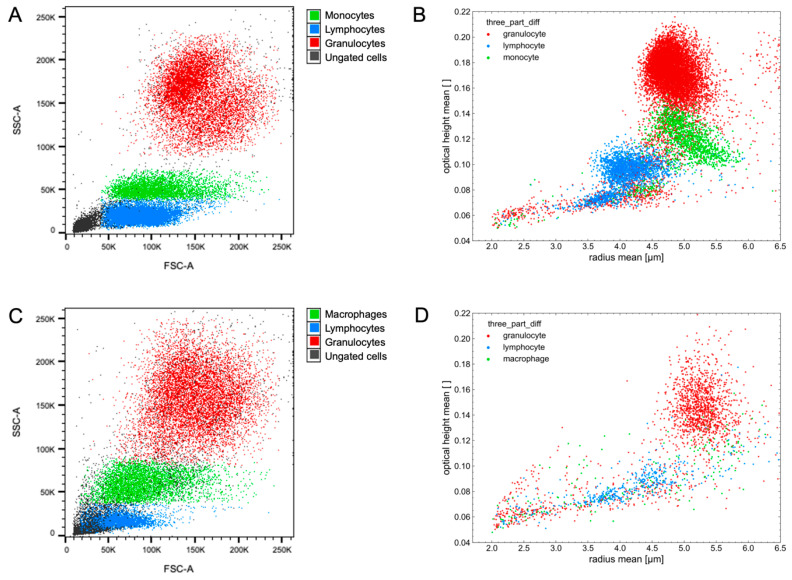
For several years, the determination of a differential cell count of a raw milk sample has been proposed as a more accurate tool for monitoring the udder health of dairy cows compared with using the absolute somatic cell count. However, the required sample preparation and staining process can be labor- and cost-intensive. Therefore, the aim of our study was to demonstrate the feasibility of analyzing unlabeled blood and milk leukocytes from dairy cows by means of digital holographic microscopy (DHM). For this, we trained three different machine learning methods, i.e., k-Nearest Neighbor, Random Forests, and Support Vector Machine, on sorted leukocyte populations (granulocytes, lymphocytes, and monocytes/macrophages) isolated from blood and milk samples of three dairy cows by using fluorescence-activated cell sorting. Afterward, those classifiers were applied to differentiate unlabeled blood and milk samples analyzed by DHM. A total of 70 blood and 70 milk samples were used. Those samples were collected from five clinically healthy cows at 14-time points within a study period of 26 days. The outcome was compared with the results of the same samples analyzed by flow cytometry and (in the case of blood samples) also to routine analysis in an external laboratory. Moreover, a standard vaccination was used as an immune stimulus during the study to check for changes in cell morphology or cell counts. When applied to isolated leukocytes, Random Forests performed best, with a specificity of 0.93 for blood and 0.84 for milk cells and a sensitivity of 0.90 and 0.81, respectively. Although the results of the three analytical methods differed, it could be demonstrated that a DHM analysis is applicable for blood and milk leukocyte samples with high reliability. Compared with the flow cytometric results, Random Forests showed an MAE of 0.11 (SD = 0.04), an RMSE of 0.13 (SD = 0.14), and an MRE of 1.00 (SD = 1.11) for all blood leukocyte counts and an MAE of 0.20 (SD = 0.11), an RMSE of 0.21 (SD = 0.11) and an MRE of 1.95 (SD = 2.17) for all milk cell populations. Further studies with larger sample sizes and varying immune cell compositions are required to establish method-specific reference ranges.
Keywords: dairy cow; differential cell count; differential leukocyte count; digital holographic microscopy; immunomonitoring; immunophenotyping; somatic cell count; udder health.
Conflict of interest statement
The authors declare that they have no conflicts of interest.
Figures


Similar articles
-
Development of an advanced flow cytometry based high-resolution immunophenotyping method to benchmark early immune response in dairy cows.Sci Rep. 2021 Nov 24;11(1):22896. doi: 10.1038/s41598-021-02405-w. Sci Rep. 2021. PMID: 34819614 Free PMC article.
-
Differential somatic cell count-A novel method for routine mastitis screening in the frame of Dairy Herd Improvement testing programs.J Dairy Sci. 2017 Jun;100(6):4926-4940. doi: 10.3168/jds.2016-12409. Epub 2017 Mar 30. J Dairy Sci. 2017. PMID: 28365116
-
Using High-Resolution Differential Cell Counts (HRDCCs) in Bovine Milk and Blood to Monitor the Immune Status over the Entire Lactation Period.Animals (Basel). 2022 May 24;12(11):1339. doi: 10.3390/ani12111339. Animals (Basel). 2022. PMID: 35681803 Free PMC article.
-
Flow cytometric differential cell counts in milk for the evaluation of inflammatory reactions in clinically healthy and subclinically infected bovine mammary glands.J Dairy Sci. 2011 Oct;94(10):5033-44. doi: 10.3168/jds.2011-4348. J Dairy Sci. 2011. PMID: 21943754
-
Advantages and Challenges of Differential Immune Cell Count Determination in Blood and Milk for Monitoring the Health and Well-Being of Dairy Cows.Vet Sci. 2022 May 27;9(6):255. doi: 10.3390/vetsci9060255. Vet Sci. 2022. PMID: 35737307 Free PMC article. Review.
References
-
- Oviedo-Boyso J., Valdez-Alarcon J.J., Cajero-Juarez M., Ochoa-Zarzosa A., Lopez-Meza J.E., Bravo-Patino A., Baizabal-Aguirre V.M. Innate immune response of bovine mammary gland to pathogenic bacteria responsible for mastitis. J. Infect. 2007;54:399–409. doi: 10.1016/j.jinf.2006.06.010. - DOI - PubMed
LinkOut - more resources
Full Text Sources
