. 2024 Oct 23;25(21):11380.
doi: 10.3390/ijms252111380.

7-Ketocholesterol Effects on Osteogenic Differentiation of Adipose Tissue-Derived Mesenchymal Stem Cells

Affiliations

7-Ketocholesterol Effects on Osteogenic Differentiation of Adipose Tissue-Derived Mesenchymal Stem Cells

Beatriz Araújo Oliveira et al. Int J Mol Sci. .

Abstract

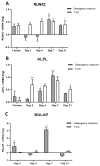
Some oxysterols were shown to promote osteogenic differentiation of mesenchymal stem cells (MSCs). Little is known about the effects of 7-ketocholesterol (7-KC) in this process. We describe its impact on human adipose tissue-derived MSC (ATMSC) osteogenic differentiation. ATMSCs were incubated with 7-KC in osteogenic or adipogenic media. Osteogenic and adipogenic differentiation was evaluated by Alizarin red and Oil Red O staining, respectively. Osteogenic (ALPL, RUNX2, BGLAP) and adipogenic markers (PPARƔ, C/EBPα) were determined by RT-PCR. Differentiation signaling pathways (SHh, Smo, Gli-3, β-catenin) were determined by indirect immunofluorescence. ATMSCs treated with 7-KC in osteogenic media stained positively for Alizarin Red. 7-KC in adipogenic media decreased the number of adipocytes. 7-KC increased ALPL and RUNX2 but not BGLAP expressions. 7-KC decreased expression of PPARƔ and C/EBPα, did not change SHh, Smo, and Gli-3 expression, and increased the expression of β-catenin. In conclusion, 7-KC favors osteogenic differentiation of ATMSCs through the expression of early osteogenic genes (matrix maturation phase) by activating the Wnt/β-catenin signaling pathway, while inhibiting adipogenic differentiation. This knowledge can be potentially useful in regenerative medicine, in treatments for bone diseases.

Keywords: 7-KC; Wnt/β-catenin; adipose tissue; mesenchymal stem cell; osteogenic differentiation; oxysterol.


Conflict of interest statement

The authors declare no conflicts of interest.

Figures

Figure 1
Figure 1
Osteogenic and adipogenic differentiation of ATMSCs. Effect of 7-KC. Osteogenic differentiation: (A): Control, ATMSCs cultivated for 21 days with osteogenic specific medium alone. (B,C): Osteogenic medium with 3 and 5 µM 7-KC, respectively. Staining with Alizarin Red. Bar scale: 12.5 µm. Adipogenic differentiation: (D): Control, ATMSCs cultivated for 14 days with adipogenic specific medium. (E,F): Adipogenic medium with 3 and 5 µM 7-KC M, respectively. Staining with Oil Red O. White bar scale: 200 µm, black bar scale: 50 µm.
Figure 2
Figure 2
Expression of osteogenic differentiation markers in ATMSCs as measured by RT-PCR. ATMSCs were incubated with osteogenic specific medium with or without 10 µM 7-KC for different periods of time. (A): RUNX2. (B): ALPL. (C): BGLAP. Data are mean ± SD from three independent experiments in duplicate. Results are shown in log scale. * p < 0.05; ** p < 0.01; *** p < 0.0001.
Figure 3
Figure 3
Expression of adipogenic differentiation markers in ATMSCs as measured by RT-PCR. ATMSCs were incubated with osteogenic specific medium with or without 10 µM 7-KC for different periods of time. (A): PPARƔ expression. (B): C/EBPα expression. ATMSCs were incubated with osteogenic or adipogenic specific medium with or without 10 µM 7-KC for 21 days. (C): PPARƔ expression. (D): C/EBPα expression. Data are mean ± SD from three independent experiments in duplicate. Results are expressed in log scale. * p < 0.05; ** p < 0.01; *** p < 0.0001.
Figure 4
Figure 4
Osteogenic signaling pathways measured by immunofluorescence. Effect of 7-KC and osteogenic and adipogenic media. (A): Mean fluorescence intensity of SHh expression. (B,C): Mean fluorescence intensity of Smo expression in membrane/cytoplasm and nucleus, respectively. (D,E): Mean fluorescence intensity of Gli-3 expression in membrane/cytoplasm and nucleus, respectively. (F): Percentage of cells expressing β-catenin in membrane/cytoplasm. (G): Mean fluorescence intensity of β-catenin expression in membrane/cytoplasm. (H): Percentage of cells expressing β-catenin in the nucleus. (I): Mean fluorescence intensity of β-catenin expression in the nucleus. Data are mean ± SD from three independent experiments in duplicate. * p < 0.05; ** p < 0.01.

References

    1. Perez-Silos V., Camacho-Morales A., Fuentes-Mera L. Mesenchymal Stem Cells Subpopulations: Application for Orthopedic Regenerative Medicine. Stem Cells Int. 2016;2016:3187491. doi: 10.1155/2016/3187491. - DOI - PMC - PubMed
    1. Smith Q., Stukalin E., Kusuma S., Gerecht S., Sun S.X. Stochasticity and Spatial Interaction Govern Stem Cell Differentiation. Dyn. Sci. Rep. 2015;5:12617. doi: 10.1038/srep12617. - DOI - PMC - PubMed
    1. Schwartz L., da Veiga Moreira J., Jolicoeur M. Physical forces modulate cell differentiation and proliferation processes. J. Cell. Mol. Med. 2018;22:738–745. doi: 10.1111/jcmm.13417. - DOI - PMC - PubMed
    1. Frank V., Kaufmann S., Wright R., Horn P., Yoshikawa H.Y., Wuchter P., Madsen J., Lewis A.L., Armes S.P., Ho A.D., et al. Frequent mechanical stress suppresses proliferation of mesenchymal stem cells from human bone marrow without loss of multipotency. Sci. Rep. 2016;6:24264. doi: 10.1038/srep24264. - DOI - PMC - PubMed
    1. Ozkul Y., Galderisi U. The Impact of Epigenetics on Mesenchymal Stem Cell Biology. J. Cell. Physiol. 2016;231:2393–2401. doi: 10.1002/jcp.25371. - DOI - PubMed

MeSH terms

LinkOut - more resources