. 2024 Oct 23;25(21):11409.
doi: 10.3390/ijms252111409.

Gestational Diabetes Mellitus-Induced Inflammation in the Placenta via IL-1β and Toll-like Receptor Pathways

Affiliations

Gestational Diabetes Mellitus-Induced Inflammation in the Placenta via IL-1β and Toll-like Receptor Pathways

Katarzyna Zgutka et al. Int J Mol Sci. .

Abstract

Gestational diabetes mellitus is characterised by an insufficient insulin response to hyperglycaemia and the development of insulin resistance. This state has adverse effects on the health outcomes of the mother and child. Existing hyperglycaemia triggers a state of inflammation that involves several tissues, including the placenta. In this study, we analysed the putative pathomechanism of GDM, with special emphasis on the role of chronic, sterile, pro-inflammatory pathways. The expression and regulation of the elements of IL-1β and Toll-like receptor (TLR) pathways in GDM maternal blood plasma, healthy placental explants and a choriocarcinoma cell line (BeWo cell line) stimulated with pro-inflammatory factors was evaluated. Our results indicate elevated expression of the IL-1β and TLR pathways in GDM patients. After stimulation with IL-1β or LPS, the placental explants and BeWo cell line showed increased production of pro-inflammatory IL-6, TNFa and IL-1β together with increased expression of the elements of the signalling pathways. The application of selected inhibitors of NF-ĸB, MAPK and recombinant interleukin 1 receptor antagonist (IL1RA) proved the key involvement of the IL-1β pathway and TLRs in the pathogenesis of GDM. Our results show the possible existence of loops of autocrine stimulation and a possible inflammatory pathomechanism in placentas affected by GDM.

Keywords: GDM; gestational diabetes mellitus; inflammation; interleukin.


Conflict of interest statement

The authors declare no conflicts of interest.

Figures
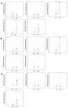
Figure 1
Figure 1
A box and whisker plot showing the absolute mRNA expression of elements of the IL-1β pathway (Panel (A)), Toll-like receptors (Panel (B)) and elements of the intracellular signal transduction pathway (Panel (C)), as assessed by RQ-PCR. The results are presented as the ratio of the gene of interest vs. internal control expression (β2-M; β2-microglobulin); * p < 0.05. The dot in the middle of the box represents the median value, and the single dots outside the box represent outliers. NGT (explants from patients with normal glucose tolerance), GDM (explants from patients with gestational diabetes mellitus), IL-1β (interleukin-1β), IL-1α (interleukin-1α), IL1R1 (interleukin 1 receptor), IL1RAP (interleukin 1 receptor accessory protein), IL1RA (interleukin-1 receptor antagonist), TLRs 1, 2, 4, 6, 10 (Toll-like receptors 1, 2, 4, 6, 10), RELA (nuclear factor NF-kappa-B p65 subunit), MYD88 (myeloid differentiation primary response 88), TRAF6 (TNF receptor-associated factor 6) and CD14 (myeloid cell-specific leucine-rich glycoprotein).
Figure 2
Figure 2
A box and whisker plot showing the concentration of IL1RA in the peripheral blood plasma of GDM and NGT women as assessed by an immunoenzymatically assay. * p < 0.05. The dot in the middle of the box represents the median value. NGT (peripheral blood from patients with normal glucose tolerance), GDM (peripheral blood from patients with gestational diabetes mellitus), IL1RA (interleukin-1 receptor antagonist).
Figure 3
Figure 3
The effects of placental tissue (left) and BeWo cell (right) stimulation by pro-inflammatory molecules, IL1-β (20 ng/mL) and LPS (100 ng/mL) on relative mRNA expression of IL-6, IL-1β and TNF-α. The data are presented as the fold difference, where 1 = unstimulated control samples. * p < 0.05 compared with unstimulated controls. All experiments were repeated at least three times with similar results. IL-6 (interleukin 6), IL-1β (interleukin 1β), TNF-α (tumour necrosis factor α), LPS (lipopolysaccharide).
Figure 4
Figure 4
The relative mRNA expression of elements of the IL-1β system (Panels (A) and (D)), Toll-like receptors (Panels (C) and (F)) and elements of intracellular signal transduction pathways (Panels (B) and (E)), as assessed by RQ-PCR in placental tissue (Panels (AC)) and BeWo cells (Panels (DF)) stimulated by LPS (100 ng/mL) or IL-1β (20 ng/mL). The data are presented as fold differences, where 1 = unstimulated controls. * p < 0.05 compared with unstimulated controls. IL-1β (interleukin 1β), IL1R1 (interleukin 1 receptor), IL1RAP (IL1R1 accessory protein), IL1RA (IL1R1 antagonist), TLRs 1, 2, 4, 6 and 10 (Toll-like Receptors 1, 2, 4, 6 and 10), RELA (nuclear factor NF-kappa-B p65 subunit), CD14 (myeloid cell-specific leucine-rich glycoprotein), MYD88 (myeloid differentiation primary response 88), TRAF6 (TNF receptor-associated factor 6) and LPS (lipopolysaccharide).
Figure 5
Figure 5
Western blot analysis of changes in protein expression after stimulation with LPS (100 ng/mL) or IL-1β (20 ng/mL). GAPDH (glyceraldehyde-3-phosphate dehydrogenase) served as the loading control. IL-1β (interleukin1β), IL1R1 (interleukin 1 receptor), IL1RAP (interleukin 1 receptor accessory protein), IL1RA (IL1R antagonist), TLRs 1, 2, 4, 6 and 10 (Toll-like receptors 1, 2, 4, 6 and 10), MYD88 (myeloid differentiation primary response 88), TRAF6 (TNF receptor-associated factor 6) and LPS (lipopolysaccharide).
Figure 6
Figure 6
Measurement of pro-inflammatory cytokine concentrations in supernatants collected from placenta explants stimulated with IL-1β (20 ng/mL) (Panel (A)) or with LPS (100 ng/mL) (Panel (B)) and with selected inhibitors: recombinant IL1RA (100 ng/mL); BMS 345541(12.5 µM)—a highly selective inhibitor of I kappa B kinase that blocks NF-κB-dependent transcription; and a MAPK (mitogen-activated protein kinase) inhibitor—PD184352 (5 µM). * p < 0.05 compared with unstimulated controls. ** p < 0.05 compared with stimulated controls and after the application of inhibitors. IL-1β (interleukin1β), IL-6 (interleukin 6), IL1R (interleukin 1 receptor), LPS (lipopolysaccharide), TNF-α (tumour necrosis factor α), IL1RA (IL1R antagonist).
Figure 7
Figure 7
Relative downregulation of mRNA expression of Toll-like receptors 1, 2, 6 and 10 (Panel (A)) and IL1RA, TRAF6 and RELA (Panel (B)) in placental explants stimulated with IL-1β alone or by IL-1β with selected inhibitors. UC (unstimulated controls), expression = 1. * p < 0.05 compared with IL-1β-stimulated samples. TLRs 1, 2, 6, 10 (Toll-like receptors 1, 2, 6, 10), IL-1β (interleukin 1β), IL1RA (IL-1R antagonist), TRAF6 (TNF receptor-associated factor 6), RELA (nuclear factor NF-kappa-B p65 subunit), BMS 345541 (a highly selective inhibitor of I kappa B kinase that blocks NF-κB-dependent transcription), PD184352 (an MAPK (mitogen-activated protein kinase) inhibitor).

References

    1. McElwain C.J., McCarthy F.P., McCarthy C.M. Gestational Diabetes Mellitus and Maternal Immune Dysregulation: What We Know So Far. Int. J. Mol. Sci. 2021;22:4261. doi: 10.3390/ijms22084261. - DOI - PMC - PubMed
    1. International Diabetes Federation. [(accessed on 7 December 2021)]. Available online: https://diabetesatlas.org/data/en/indicators/14/
    1. Nguyen-Ngo C., Jayabalan N., Salomon C., Lappas M. Molecular pathways disrupted by gestational diabetes mellitus. J. Mol. Endocrinol. 2019;63:R51–R72. doi: 10.1530/JME-18-0274. - DOI - PubMed
    1. Baeyens L., Hindi S., Sorenson R.L., German M.S. Β-cell adaptation in pregnancy. Diabetes Obes. Metab. 2016;18:63–70. doi: 10.1111/dom.12716. - DOI - PMC - PubMed
    1. Maedler K., Sergeev P., Ris F., Oberholzer J., Joller-Jemelka H.I., Spinas G.A., Kaiser N., Halban P.A., Donath M.Y. Glucoseinduced beta cell production of IL-1beta contributes to glucotoxicity in human pancreatic islets. J. Clin. Investig. 2002;110:851–860. doi: 10.1172/JCI200215318. - DOI - PMC - PubMed

LinkOut - more resources