. 2024 Oct 24;25(21):11423.
doi: 10.3390/ijms252111423.

NRXN1 as a Prognostic Biomarker: Linking Copy Number Variation to EMT and Survival in Colon Cancer

Affiliations

NRXN1 as a Prognostic Biomarker: Linking Copy Number Variation to EMT and Survival in Colon Cancer

Hyun Jin Bang et al. Int J Mol Sci. .

Abstract

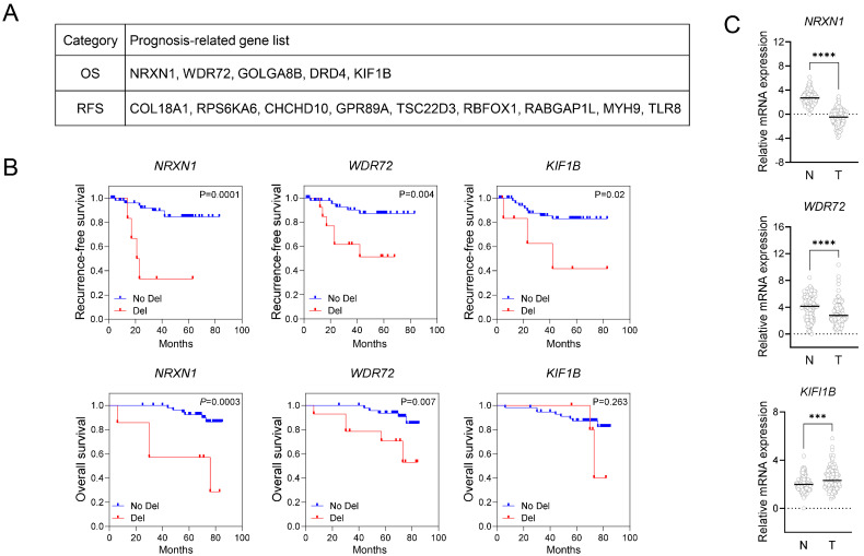
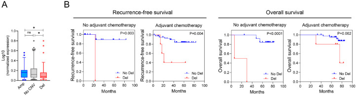
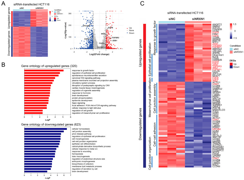
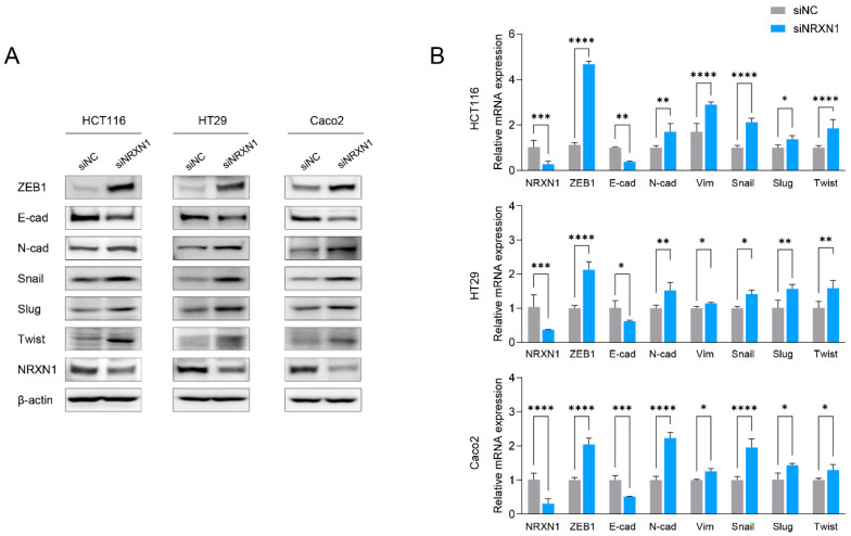
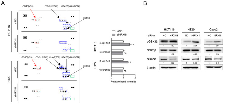
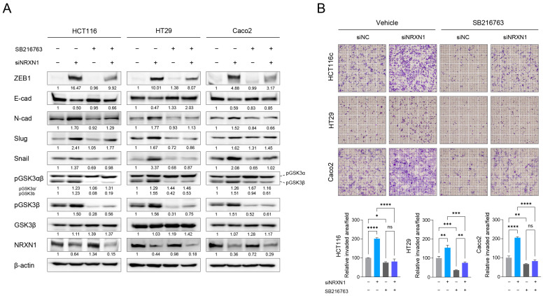
The role of biomarkers in cancer treatment varies significantly depending on the cancer stage. Thus, in clinical practice, tailoring biomarkers to meet the specific needs and challenges of each cancer stage can increase the precision of treatment. Because they reflect underlying genetic alterations that influence cancer progression, copy number variation (CNV) biomarkers can play crucial prognostic roles. In our previous study, we identified potential survival-related genes for colorectal cancer (CRC) by analyzing CNV and gene expression data using a machine-learning approach. To further investigate the biological function of NRXN1, we assessed the use of RNA sequencing, phosphokinase assays, real-time quantitative PCR, and Western blot analysis. We found that NRXN1 copy number deletion was significantly associated with poor overall survival (OS) and recurrence-free survival (RFS), even in patients who received adjuvant chemotherapy. Compared with its expression in normal tissues, NRXN1 expression was lower in tumors, suggesting its potential role as a tumor suppressor. NRXN1 knockdown enhanced CRC cell viability and invasion, and transcriptome analysis indicated that the increased invasion was caused by GSK3β-mediated epithelial-mesenchymal transition. These findings highlight NRXN1 copy number deletion as a novel biomarker for predicting recurrence and survival in patients with resected colon cancer.

Keywords: colorectal cancer (CRC); epithelial–mesenchymal transition; mechanisms of inhibition.


Conflict of interest statement

The authors declare no conflicts of interest.

Figures

Figure 1
Figure 1
Identification of survival-related candidate genes in colorectal cancer. (A) Gene list of putative survival-related factors identified by ML analysis based on integrating CNV and gene expression data. (B) Predictive values of putative survival-related genes for CRC patients. Left: Kaplan–Meier curves for CNVs of the indicated genes associated with RFS and OC in CRC patients (stage III, n = 63). Kaplan–Meier plots were constructed based on the copy number status of each gene to determine the difference between cases with gene-harboring copy number deletion versus unaltered status. Blue indicates no deletion; red indicates copy number deletion. The log-rank p-value for significance between the curves is indicated at the top of each panel within the figure. (C) Differential gene expression analysis of NRXN1, WDR72, and KIF1B in tumor and normal tissues (stage II, III, n = 120). qRT-PCR determined the gene expression of the indicated genes. The expression of GAPDH was used as an internal reference control for qRT-PCR analysis. OS, overall survival; RFS, Recurrence-free survival. Data are presented as mean ± SD. *** p < 0.001, **** p < 0.0001.
Figure 2
Figure 2
The correlation between NRXN1 CNV and gene expression, and Kaplan–Meier survival analyses based on NRXN1 CNV after adjuvant chemotherapy. (A) The correlation between CNV (in genomic DNA) and gene expression (in mRNA) of NRXN1 in CRC (stage II, III; n = 120). qRT-PCR determined NRXN1 expression. Expression of GAPDH was used as an internal reference control for RT-qPCR analysis. No CNV, no copy number variation; Amp, amplification; Del, deletion. (B) Kaplan–Meier plots for NRXN1 CND associated with the recurrence-free survival and overall survival of CRC patients with or without adjuvant chemotherapy, including 5-fluorouracil and oxaliplatin. n indicates the number of cases and events. Blue indicates no CND; red indicates NRXN1 CND. The log-rank p-value for significance between the curves is indicated at the bottom of each panel. NRXN1 CND shows a significant association with poor RFS and OS in the adjuvant chemotherapy receiving group. Data are presented as mean ± SD. ns; no significant, * p < 0.05.
Figure 3
Figure 3
NRXN1 knockdown enhances CRC cell viability and significantly increases invasion. Comparison of cell viability (A) and invasion potential (B) of CRC cell lines transfected with siNC or siNRXN1. Western blot analysis verifying NRXN1 protein knockdown in siNRXN1-transfected cells. Data are presented as mean ± SD. *** p < 0.001, **** p < 0.0001.
Figure 4
Figure 4
Transcriptome analysis of NRXN1 knockdown cells. (A) Heatmap and volcano plots showing the DEGs in siNRXN1-treated HCT116 cells compared to siNC (n = 3/group). (B) GO enrichment analysis of the biological process terms associated with 320 upregulated and 623 downregulated genes. (C) Heatmap depicting the gene expression related to the response to growth factor and regulation of epithelial cell proliferation, mesenchymal cell proliferation, cell junction assembly, cell–cell adhesion, and cell junction organization. Genes in red indicate those regulated by NRXN1 knockdown and associated with Epithelial-Mesenchymal Transition (EMT). DEG; differentially expressed genes, siNC; negative control siRNA, siNRXN1; NRXN1 siRNA.
Figure 5
Figure 5
NRXN1 knockdown induces EMT in CRC cells. Effect of siRNA-mediated NRXN1 gene silencing on the EMT signaling pathway in CRC cells. (A) Gene expression of the EMT-related signaling pathway was determined using Western blot analysis with the indicated antibodies. β-actin was used as the loading control. (B) qRT-PCR determined the mRNA expression of EMT-related genes. siNC; negative control siRNA, siNRXN1; NRXN1 siRNA. Data are presented as mean ± SD. * p < 0.05, ** p < 0.01, *** p < 0.001, **** p < 0.0001.
Figure 6
Figure 6
NRXN1 knockdown induces GSK3β phosphorylation. (A) Left: the images of phospho-kinase arrays with marked proteins. Positive reference double spots are marked in black rectangles; the area of negative control spots is marked in green rectangles. Phosphorylated kinase spots showing the difference between siNC and siNRXN1 are marked with blue and red rectangles. Blue indicates kinases that are not commonly altered between HCT116 and HT29, while red indicates those that are consistently altered in both cell lines. Right: bar graph showing the relative change in Ser9 phospho-GSK3β and positive reference spot intensity between siNC- and siNRXN1-transfected cells. Spot intensities were quantified using ImageJ software (version 1.53k) and normalized to those of positive controls on the same membrane. Data are presented as mean ± SD. ns; not significant, * p < 0.05 (B) Western blot analysis of GSK3β phosphorylation at its residue Ser9. β-actin was used as the loading control. Band intensity quantification is labeled below the blot.
Figure 7
Figure 7
NRXN1 knockdown promotes the invasion of CRC cell through GSK3β-mediated EMT induction. The effect of GSK3β inhibition on NRXN1 knockdown-induced EMT and invasion property in CRC. siNC or siNRXN1-transfected HCT116, HT29, and Caco2 cells were treated with GSK3β inhibitor, SB216763 (10 µM) for 24 h. (A) The EMT signaling pathway was evaluated by Western blot analysis with the indicated antibodies. β-actin was used as the loading control. Band intensity quantification is labeled below the blot. (B) Upper: representative Transwell invasion assay of siRNA-transfected CRC cells treated with vehicle or SB216763 (10 µM). Lower: quantification of invasive capacities of CRC cells. Data are presented as mean ± SD. ns; no significant, * p < 0.05, ** p < 0.01, *** p < 0.001, **** p < 0.0001.

Similar articles

References

    1. Benson A.B., III, Adam M., Chang G.J., Chen Y.-J., Ciombor K.K., Cohen S.A., Cooper H.S., Deming D. Colon cancer, version 3.2024, NCCN Clinical Practice Guidelines in Oncology. Natl. Compr. Cancer Netw. 2024;22:e240029. doi: 10.6004/jnccn.2024.0029. - DOI - PubMed
    1. André T., Boni C., Navarro M., Tabernero J., Hickish T., Topham C., Bonetti A., Clingan P., Bridgewater J., Rivera F., et al. Improved overall survival with oxaliplatin, fluorouracil, and leucovorin as adjuvant treatment in stage II or III colon cancer in the MOSAIC trial. J. Clin. Oncol. 2009;27:3109–3116. doi: 10.1200/JCO.2008.20.6771. - DOI - PubMed
    1. Kotani D., Oki E., Nakamura Y., Yukami H., Mishima S., Bando H., Shirasu H., Yamazaki K., Watanabe J., Kotaka M., et al. Molecular residual disease and efficacy of adjuvant chemotherapy in patients with colorectal cancer. Nat. Med. 2023;29:127–134. doi: 10.1038/s41591-022-02115-4. - DOI - PMC - PubMed
    1. Ogunwobi O.O., Mahmood F., Akingboye A. Biomarkers in Colorectal Cancer: Current Research and Future Prospects. Int. J. Mol. Sci. 2020;21:5311. doi: 10.3390/ijms21155311. - DOI - PMC - PubMed
    1. Redon R., Ishikawa S., Fitch K.R., Feuk L., Perry G.H., Andrews T.D., Fiegler H., Shapero M.H., Carson A.R., Chen W., et al. Global variation in copy number in the human genome. Nature. 2006;444:444–454. doi: 10.1038/nature05329. - DOI - PMC - PubMed

MeSH terms

Substances

LinkOut - more resources