Review
. 2024 Oct 24;25(21):11429.
doi: 10.3390/ijms252111429.

Linking Environmental Genotoxins to Neurodegenerative Diseases Through Transcriptional Mutagenesis

Affiliations
Review

Linking Environmental Genotoxins to Neurodegenerative Diseases Through Transcriptional Mutagenesis

Bert M Verheijen et al. Int J Mol Sci. .

Abstract

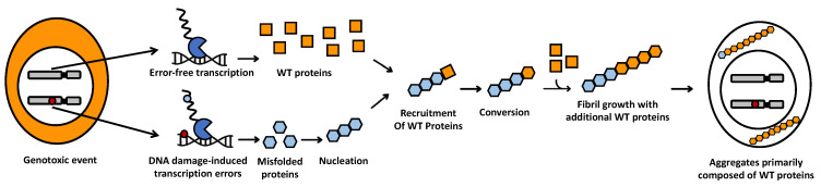
Numerous lines of evidence suggest that DNA damage contributes to the initiation, progression, and severity of neurodegenerative diseases. However, the molecular mechanisms responsible for this relationship remain unclear. This review integrates historical data with contemporary findings to propose that DNA damage exacerbates neurodegenerative diseases by inducing transcription errors. First, we describe the scientific rationale and basic biological concepts that underpin this hypothesis. Then, we provide epidemiological, cellular, and molecular data to support this idea, and we describe new and recently published observations that suggest that the former high incidence of neurodegenerative disease in Guam may have been driven by DNA damage-induced transcription errors. Finally, we explore the long-term implications of these findings on our understanding of the impact of genotoxic stress on human aging and disease.

Keywords: DNA damage; mutagenesis; mutant proteins; neurodegenerative diseases; protein aggregation; transcription errors.


Conflict of interest statement

The authors declare no conflict of interest.

Figures

Figure 1
Figure 1
Mechanism proposed to give rise to the protein aggregates characteristic of amyloid- and prion-like diseases. After a genotoxic event, transcription errors (small blue circle) are made on damaged DNA templates (small red circle) by RNA polymerases (blue “Pacman”). These errors give rise to a limited number of mutant proteins with enhanced amyloid- or prion-like properties (blue honeycombs) that convert WT proteins (orange squares) to an amyloid- or prion-like shape. Converted WT proteins (orange honeycombs) then recruit additional WT proteins to the amyloid seed to perpetuate fibril growth without the need for further errors.
Figure 2
Figure 2
Transcription errors give rise to proteins with increased amyloid behavior. (A) A transcription error (Tr) was identified in the SOD1 transcript that mimics a mutation (Mut) implicated in amyotrophic lateral sclerosis. This error substituted a guanine for an adenine base, resulting in substitution of a glycine (G) for a glutamic acid residue (E) in the protein. (B) SOD1 is soluble and present throughout the cell, including the nucleus. (C) In contrast, SOD1 proteins made from erroneous transcripts form aggregates that excluded the nucleus. (D) Quantification of SOD1 aggregation and mislocalization. * p < 0.05; ** p < 0.01. (EG) When WT and transcript error (TE)-derived SOD1 are expressed simultaneously; erroneous SOD1 corrupts WT SOD1 and recruits it into extranuclear aggregates. (H) Quantification of SOD1 colocalization with N-terminal or C-terminal tags. Scale bars 30 μm (B,C); 20 μm (EG). Figure adapted from [29].
Figure 3
Figure 3
Methylazoxymethanol (MAM) induces transcriptional mutagenesis in cultured cells. (A) Schematic diagram of the experiment. Primary mouse hippocampal neural stem cells (NSCs) were grown in BMP4+/EGF− culturing medium for 3 days to put them in a quiescent state. Next, cells were treated with a high dose of MAM acetate (1 mM) or vehicle (phosphate-buffered saline [PBS]) for 1 h, after which they were extensively rinsed with PBS and cultured for 16 h. Cells were then collected and used immediately for single-cell RNA sequencing (scRNA-seq) experiments (10x Genomics v3.1). All cells used for experiments described in [31] were cultured at 37 °C under 5% CO2 and 5% O2. Following quiescence induction, cells were kept in BMP4+/EGF− culturing medium for treatments and 16 h intervals. The photomicrograph shows representative adherent NSCs in culture. Scale bar: 75 µm. (B) scRNA-seq data were processed, and individual cells were depicted in Uniform Manifold Approximation and Projection (UMAP) plots. (C,D) A transcriptionally distinct cluster of cells associated with cell proliferation genes (e.g., Top2a and Mki67) was observed. The dot plot depicts the expression of marker genes in each cluster. The diameter of the dots indicates the percentage of cells expressing the genes. This clustering was used to estimate the proportions of proliferating and arrested cells. (E) Bar plots depict the proportions of each cluster for each condition, which indicates that the majority of cells in the experiment were arrested. (F) Transcriptional error spectra in NSCs following exposure to MAM. MAM-treated NSCs showed an increased C→U error rate (average error rate: 6.9 × 10⁻⁵/bp) as compared to vehicle (PBS)-treated cells (average error rate: 6.7 × 10⁻⁶/bp). C→U errors correspond to transcriptional mutagenesis on O6-meG DNA lesions induced by MAM. (G) Analysis of pseudo-alleles (transcripts containing an error at an identical sequence location) revealed that a substantial number of repeated transcription errors occurred in MAM-treated cells (analysis of all MAM-treated cells combined). Only alleles with more than 10% mutant RNA were included. Numbers above bars indicate number of pseudo-alleles detected. (H) Comparison of arrested MAM-treated cells and arrested control (vehicle-treated) cells indicated that expression of transcriptional markers for autophagy induction [54] was upregulated in MAM-treated cells, although most of these changes were not statistically significant (pseudobulk limma-voom workflow). Source data were from [31] (note that in source the notation [expected sequencing read: A/C/G/T] → [interpretation of observed read: A/C/G/U] was used for transcript error spectra). Abbreviations aNSC: activated NSC; BMP4: bone morphogenetic protein 4; EGF: epidermal growth factor; FGF: fibroblast growth factor-basic; qNSC: quiescent NSC.

Similar articles

Cited by

References

    1. Cannon J.R., Greenamyre J.T. The role of environmental exposures in neurodegeneration and neurodegenerative diseases. Toxicol. Sci. 2011;124:225–250. doi: 10.1093/toxsci/kfr239. - DOI - PMC - PubMed
    1. Sakowski S.A., Koubek E.J., Chen K.S., Goutman S.A., Feldman E.L. Role of the Exposome in Neurodegenerative Disease: Recent Insights and Future Directions. Ann. Neurol. 2024;95:635–652. doi: 10.1002/ana.26897. - DOI - PMC - PubMed
    1. Kisby G.E., Wilson D.M., 3rd, Spencer P.S. Introducing the Role of Genotoxicity in Neurodegenerative Diseases and Neuropsychiatric Disorders. Int. J. Mol. Sci. 2024;25:7221. doi: 10.3390/ijms25137221. - DOI - PMC - PubMed
    1. Knopman D.S., Amieva H., Petersen R.C., Chetelat G., Holtzman D.M., Hyman B.T., Nixon R.A., Jones D.T. Alzheimer disease. Nat. Rev. Dis. Primers. 2021;7:33. doi: 10.1038/s41572-021-00269-y. - DOI - PMC - PubMed
    1. Spillantini M.G., Crowther R.A., Jakes R., Hasegawa M., Goedert M. alpha-Synuclein in filamentous inclusions of Lewy bodies from Parkinson’s disease and dementia with lewy bodies. Proc. Natl. Acad. Sci. USA. 1998;95:6469–6473. doi: 10.1073/pnas.95.11.6469. - DOI - PMC - PubMed

LinkOut - more resources