. 2024 Oct 24;25(21):11441.
doi: 10.3390/ijms252111441.

Drugs That Induce Gingival Overgrowth Drive the Pro-Inflammatory Polarization of Macrophages In Vitro

Affiliations

Drugs That Induce Gingival Overgrowth Drive the Pro-Inflammatory Polarization of Macrophages In Vitro

Annalisa Palmieri et al. Int J Mol Sci. .

Abstract

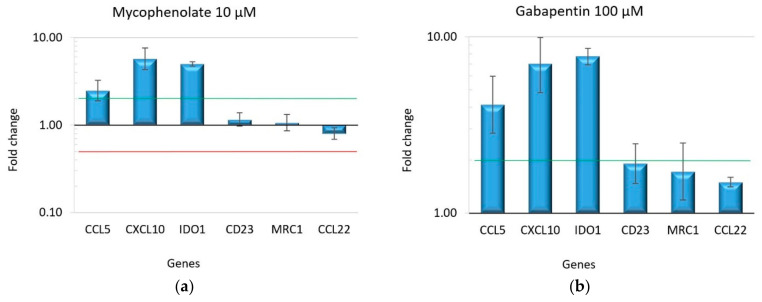
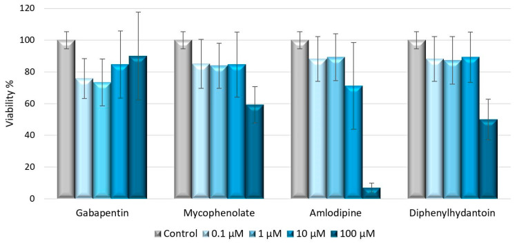
Several attempts have been made to elucidate the pathogenesis of drug-induced gingival overgrowth (DIGO), which is triggered by the chronic use of certain drugs that fall into three main categories: anticonvulsants, immunosuppressants, and calcium channel blockers. Previous research suggests that cytokines and impaired cellular functions play a role in DIGO. Of particular interest are macrophages, immune cells that can switch between M1 (pro-inflammatory) and M2 (anti-inflammatory) phenotypes in response to exogenous signals and stimuli. An imbalance between M1 and M2 macrophage populations may underlie DIGO. M1 may contribute to the initial tissue damage in DIGO, while M2 may then attempt to repair the damage with anti-inflammatory mechanisms. To test the hypothesis that drugs associated with DIGO could influence macrophage polarization, human monocytes (precursors of macrophages) were induced to differentiate into M0-naïve macrophages and then exposed to drugs: diphenylhydantoin, gabapentin, mycophenolate, and amlodipine. Quantitative real-time PCR amplification was used to measure the expression of specific genes associated with macrophage polarization. All of the drugs tested induced M0 macrophages to overexpress genes typical of the M1 phenotype, such as CCL5, CXCL10, and IDO1. This investigation provides the first evidence of a link between drugs that cause DIGO and M1 pro-inflammatory macrophage polarization. The knowledge gained from this research could be valuable for future DIGO treatment strategies.

Keywords: DIGO; gingival hyperplasia; inflammation; macrophage polarization.


Conflict of interest statement

The authors declare that they have no known competing financial interests or personal relationships that could have appeared to influence the work reported in this paper. The materials presented, and views expressed herein are the responsibility of the authors only. The sponsor takes no responsibility for any use of the information presented herein.

Figures

Figure 1
Figure 1
Immunofluorescence staining of the adhered macrophages after differentiation. Magnification 20X. (a,b) M0 macrophages negative for CD80 and CD163, respectively; (c) M1 macrophages positive for CD80 (red); (d) M1 macrophages negative for CD163; (e) M2 macrophages positive for CD163 (green); (f) M2 macrophages negative for CD80. Nuclei were stained with DAPI (blue).
Figure 2
Figure 2
Gene expression profile in differentiated macrophage subtypes M1 and M2: (a) macrophages treated with LPS and IFNγ showed the overexpression of the M1 markers and downregulation of the M2 markers; (b) IL-4 treatment induces the overexpression of the M2 markers. The bars in the graph represent the fold change in gene expression on a logarithmic scale. The error bars indicate the standard deviation of fold changes calculated from two biological samples and three experimental replicates. The green line marks a fold change = 2; the red line marks a fold change = 0.5.
Figure 3
Figure 3
The gene expression profile of macrophages treated with different drugs after 48 h. Panel (a) shows the results of gabapentin treatment, panel (b) of mycophenolate, panel (c) of amlodipine, and panel (d) of diphenylhydantoin. All the treatments induced M1 polarization, as evidenced by the significant upregulation of CCL5, CXCL10, and IDO1 genes. The bars in the graph represent the fold change in gene expression on a logarithmic scale. The error bars indicate the standard deviation of fold changes calculated from two biological samples and three experimental replications. The green line marks a fold change = 2; the red line marks a fold change = 0.5.
Figure 3
Figure 3
The gene expression profile of macrophages treated with different drugs after 48 h. Panel (a) shows the results of gabapentin treatment, panel (b) of mycophenolate, panel (c) of amlodipine, and panel (d) of diphenylhydantoin. All the treatments induced M1 polarization, as evidenced by the significant upregulation of CCL5, CXCL10, and IDO1 genes. The bars in the graph represent the fold change in gene expression on a logarithmic scale. The error bars indicate the standard deviation of fold changes calculated from two biological samples and three experimental replications. The green line marks a fold change = 2; the red line marks a fold change = 0.5.
Figure 4
Figure 4
Cell viability of macrophages (M0) treated for 48 h with different concentrations of drugs, assessed by PrestoBlue™ reagent protocol. The viability of treated samples was normalized to untreated control; error bars represent standard errors calculated from three experimental replications.

References

    1. Kimball O.P. The Treatment of Epilepsy with Sodium Diphenyl Hydantoinate. J. Am. Med. Assoc. 1939;112:1244–1245. doi: 10.1001/jama.1939.02800130028009. - DOI
    1. Drozdzik A., Drozdzik M. Drug-Induced Gingival Overgrowth-Molecular Aspects of Drug Actions. Int. J. Mol. Sci. 2023;24:5448. doi: 10.3390/ijms24065448. - DOI - PMC - PubMed
    1. Wang Q., Ji C., Smith P., McCulloch C.A. Impact of TRP Channels on Extracellular Matrix Remodeling: Focus on TRPV4 and Collagen. Int. J. Mol. Sci. 2024;25:3566. doi: 10.3390/ijms25073566. - DOI - PMC - PubMed
    1. Sume S.S., Kantarci A., Lee A., Hasturk H., Trackman P.C. Epithelial to mesenchymal transition in gingival overgrowth. Am. J. Pathol. 2010;177:208–218. doi: 10.2353/ajpath.2010.090952. - DOI - PMC - PubMed
    1. Chen P.H., Chuang Y.T., Huang C.F., Lu H.K. Expression of epithelial-mesenchymal transition-associated proteins and proliferating cell nuclear antigen in dihydropyridine-induced gingival overgrowth fibroblasts: A preliminary study. J. Dent. Sci. 2023;18:551–559. doi: 10.1016/j.jds.2022.08.025. - DOI - PMC - PubMed

MeSH terms

LinkOut - more resources