

Super-Resolution Microscopy as a Versatile Tool in Probing Molecular Assembly
- PMID: 39519049
- PMCID: PMC11545975
- DOI: 10.3390/ijms252111497
Super-Resolution Microscopy as a Versatile Tool in Probing Molecular Assembly
Abstract
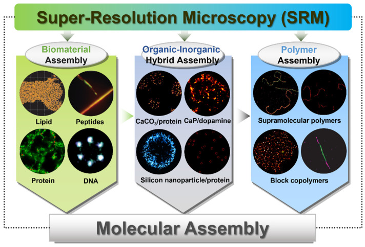
Molecular assembly is promising in the construction of advanced materials, obtaining structures with specific functions. In-depth investigation of the relationships between the formation, dynamics, structure, and functionality of the specific molecular assemblies is one of the greatest challenges in nanotechnology and chemistry, which is essential in the rational design and development of functional materials for a variety of applications. Super-resolution microscopy (SRM) has been used as a versatile tool for investigating and elucidating the structures of individual molecular assemblies with its nanometric resolution, multicolor ability, and minimal invasiveness, which are also complementary to conventional optical or electronic techniques that provide the direct observation. In this review, we will provide an overview of the representative studies that utilize SRM to probe molecular assemblies, mainly focusing on the imaging of biomolecular assemblies (lipid-based, peptide-based, protein-based, and DNA-based), organic-inorganic hybrid assemblies, and polymer assemblies. This review will provide guidelines for the evaluation of the dynamics of molecular assemblies, assembly and disassembly processes with distinct dynamic behaviors, and multicomponent assembly through the application of these advanced imaging techniques. We believe that this review will inspire new ideas and propel the development of structural analyses of molecular assemblies to promote the exploitation of new-generation functional materials.
Keywords: dynamics; interactions; molecular assembly; super-resolution microscopy.
Conflict of interest statement
The authors declare no conflicts of interest.
Figures
Similar articles
-
Probing Peptide Assembly and Interaction via High-Resolution Imaging Techniques: A Mini Review.Int J Mol Sci. 2025 Apr 23;26(9):3998. doi: 10.3390/ijms26093998. Int J Mol Sci. 2025. PMID: 40362238 Free PMC article. Review.
-
Microscopic Imaging Techniques for Molecular Assemblies: Electron, Atomic Force, and Confocal Microscopies.Chem Rev. 2021 Nov 24;121(22):14281-14347. doi: 10.1021/acs.chemrev.0c01334. Epub 2021 May 4. Chem Rev. 2021. PMID: 33942610 Review.
-
The power of super-resolution microscopy in modern biomedical science.Adv Colloid Interface Sci. 2023 Apr;314:102880. doi: 10.1016/j.cis.2023.102880. Epub 2023 Mar 13. Adv Colloid Interface Sci. 2023. PMID: 36965225 Review.
-
Super-Resolution Microscopy: From Single Molecules to Supramolecular Assemblies.Trends Cell Biol. 2015 Dec;25(12):730-748. doi: 10.1016/j.tcb.2015.10.004. Epub 2015 Nov 3. Trends Cell Biol. 2015. PMID: 26546293 Review.
-
Dynamic nanoparticle assemblies.Acc Chem Res. 2012 Nov 20;45(11):1916-26. doi: 10.1021/ar200305f. Epub 2012 Mar 26. Acc Chem Res. 2012. PMID: 22449243 Free PMC article.
Cited by
-
Probing Peptide Assembly and Interaction via High-Resolution Imaging Techniques: A Mini Review.Int J Mol Sci. 2025 Apr 23;26(9):3998. doi: 10.3390/ijms26093998. Int J Mol Sci. 2025. PMID: 40362238 Free PMC article. Review.
References
-
- Gu J., Li Z., Li Q. From single molecule to molecular aggregation science. Coord. Chem. Rev. 2023;475:214872. doi: 10.1016/j.ccr.2022.214872. - DOI
-
- Wang T., Fei J., Yu F., Xu X., Cui Y., Li J. Nanoarchitectonics of vesicle microreactors for oscillating ATP synthesis and hydrolysis. Angew. Chem. Int. Ed. 2024:e202411981. - PubMed
Publication types
MeSH terms
Substances
Grants and funding
LinkOut - more resources
Full Text Sources
