Review
. 2024 Oct 26;25(21):11506.
doi: 10.3390/ijms252111506.

Advancements in LAMP-Based Diagnostics: Emerging Techniques and Applications in Viral Detection with a Focus on Herpesviruses in Transplant Patient Management

Affiliations
Review

Advancements in LAMP-Based Diagnostics: Emerging Techniques and Applications in Viral Detection with a Focus on Herpesviruses in Transplant Patient Management

Ana Cláudia Martins Braga Gomes Torres et al. Int J Mol Sci. .

Abstract

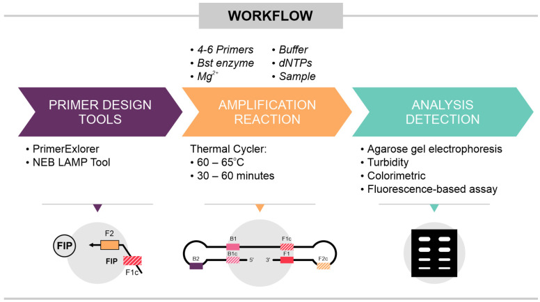
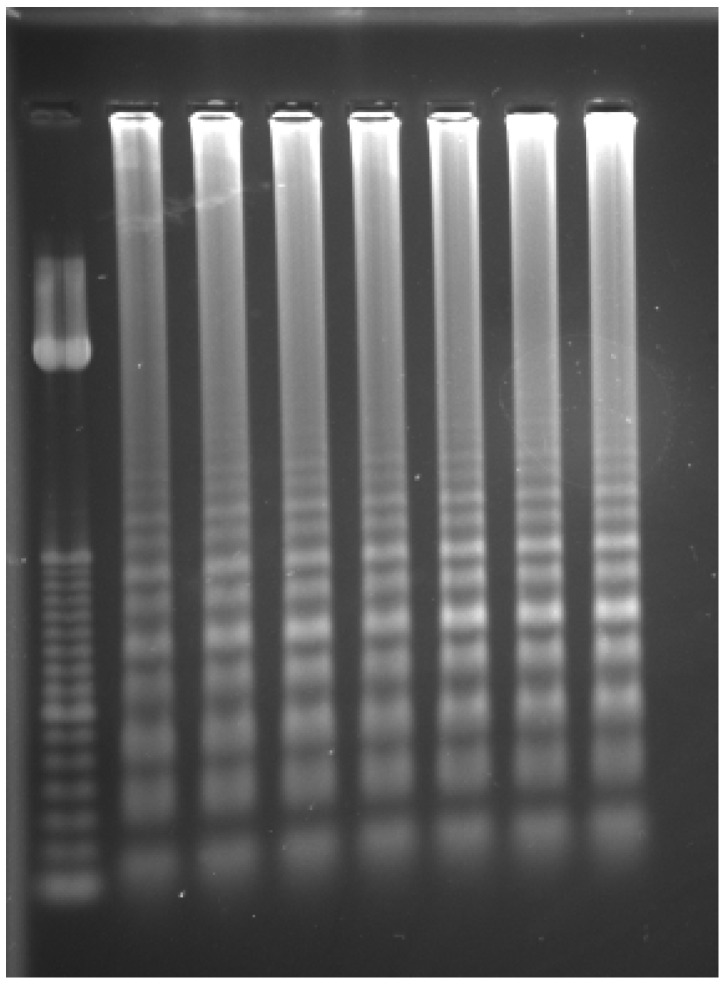
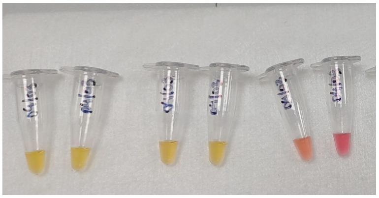
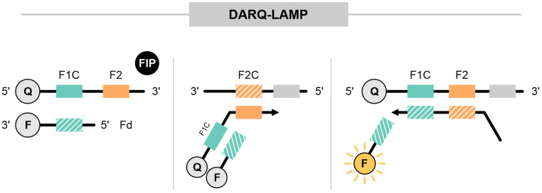
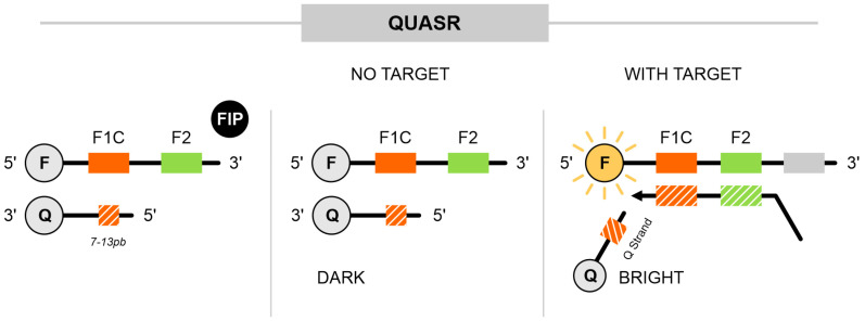
Loop-mediated isothermal amplification (LAMP) is a highly effective molecular diagnostic technique, particularly advantageous for point-of-care (POC) settings. In recent years, LAMP has expanded to include various adaptations such as DARQ-LAMP, QUASR, FLOS-LAMP, displacement probes and molecular beacons. These methods enable multiplex detection of multiple targets in a single reaction, enhancing cost-effectiveness and diagnostic efficiency. Consequently, LAMP has gained significant traction in diagnosing diverse viruses, notably during the COVID-19 pandemic. However, its application for detecting Herpesviridae remains relatively unexplored. This group of viruses is of particular interest due to their latency and potential reactivation, crucial for immunocompromised patients, including organ and hematopoietic stem cell transplant recipients. This review highlights recent advancements in LAMP for virus diagnosis and explores current research trends and future prospects, emphasizing the detection challenges posed by Herpesviridae.

Keywords: Herpesviridae; LAMP; isothermal amplification; point-of-care; viral detection.


Conflict of interest statement

The authors declare no conflicts of interest.

Figures

Figure 1
Figure 1
Diagram of loop-mediated isothermal amplification (LAMP) reaction. In a traditional LAMP assay, 4 to 6 primers are included, comprising outer primers (F3 and B3) and loop primers (LPF and LPB). Initially, FIP and F3 attach to the target sequence and initiate DNA strand synthesis, facilitated by Bst polymerase. Following this, BIP and B3 bind to their complementary targets and synthesize DNA on the opposite strands. The amplicons, with their complementary regions, undergo self-hybridization, leading to the formation of characteristic dumbbell-shaped structures.
Figure 2
Figure 2
Illustrates the workflow of the LAMP reaction, highlighting three key points. Primer Design: primers must be carefully designed to ensure specificity for the target sequence. Optimization of the LAMP Reaction: the reaction requires optimization of several parameters, including primer concentrations, the temperature for Bst (Bacillus stearothermophilus) polymerase activity, and the amplification time. Detection Methods: detection of LAMP products can be carried out in various ways, such as visualization on an agarose gel, detection of turbidity due to the release of pyrophosphate, colorimetric detection via color change (e.g., phenol red), or fluorescence-based detection using labeled probe.
Figure 3
Figure 3
Electrophoresis detection of LAMP. Illustration showing the differences between a molecular weight ladder and LAMP amplicon patterns. Left: molecular weight-size marker (ladder) used to estimate amplicon sizes. Right: gel showing seven LAMP amplicon samples.
Figure 4
Figure 4
Colorimetric method. An example of the naked-eye visualization of color changes with a phenol red pH indicator. During the amplification process, protons are released into the solution, resulting in a pH decrease. Thus, in positive reactions, the color of the reaction solution turns yellow (left), while in negative reactions it remains pink (right).
Figure 5
Figure 5
Assimilation probe (displacement probe). Primers are customized with a universal sequence (F strand) and a fluorophore attached at the 5′ end. An oligonucleotide containing a quencher is complementary to the universal sequence (Q strand). Prior to the start of the reaction, the probe and the Q strand remain bound. As the amplification begins and Bst strand displacement occurs, the quencher strand is released, resulting in signal amplification. The gray blocks represent the surrounding regions of the oligonucleotide target, while the pink and blue dotted blocks indicate regions complementary to the oligonucleotide target.
Figure 6
Figure 6
Detection of amplification by releasing of quenching (DARQ-LAMP). The FIP was modified with the attachment of a quencher molecule at the 5′ end, while a probe complementary to the F1c region included a fluorophore at the 3′ end. The modified FIP and probe remained bound. During Bst activity, a new strand was synthesized, leading to the release of the probe and detection of the signal. The gray blocks represent the surrounding regions of the oligonucleotide target, while the green and orange dotted blocks indicate regions complementary to the oligonucleotide target.
Figure 7
Figure 7
Quenching of unincorporated amplification signal reporters (QUASR). This approach includes a fluorophore at the 5′ end of either the FIP or BIP and incorporates a probe with a quencher at the 3′ end. The quencher probe is complementary to F1c only within a range of 7 to 13 base pairs, has a melting temperature approximately 10 °C lower than the LAMP reaction temperature (65 °C), and is included in the reaction at a higher concentration than the labeled primers. Thus, during amplification, primers and quencher probes remain in an unbound state. After the reaction, when the tubes are cooled, free primers and probes are in close proximity, resulting in no signal. However, when amplification occurs, primers are bound to the target, allowing the release of the signal and its detection. The gray blocks represent the surrounding regions of the oligonucleotide target, while the green and orange dotted blocks indicate regions complementary to the oligonucleotide target.
Figure 8
Figure 8
Fluorescence of loop primer upon a self-dequenching LAMP (FLOS-LAMP). In FLOS-LAMP, a fluorophore is attached to a T residue of one of the primers, without the need for a quencher. For adequate self-dequenching, the T residue chosen for fluorophore attachment must be adjacent to C (cytosine) or G (guanine) bases and other criteria. Here, we demonstrate the attachment in the LPB primer. When unbound, a self-quenching system inhibits signal release. During amplification, the labeled primer is incorporated into the reaction, leading to dequenching and the release of fluorescence.
Figure 9
Figure 9
Molecular beacons. These primers are labeled with a fluorophore at the 5′ end and a quencher molecule at the 3′ end, forming a hairpin structure. In the absence of the target, the fluorophore and quencher remain in close proximity, preventing signal emission. During the amplification process, the target binding separates the quencher and fluorophore, resulting in fluorescence detection. The light green blocks represent regions complementary within the oligonucleotide, while the solid dark green block indicates the region of the oligonucleotide that is complementary to the target region.

Similar articles

References

    1. Soroka M., Wasowicz B., Rymaszewska A. Loop-Mediated Isothermal Amplification (LAMP): The Better Sibling of PCR? Cells. 2021;10:1931. doi: 10.3390/cells10081931. - DOI - PMC - PubMed
    1. Glökler J., Lim T.S., Ida J., Frohme M. Isothermal amplifications—A comprehensive review on current methods. Crit. Rev. Biochem. Mol. Biol. 2021;56:543–586. doi: 10.1080/10409238.2021.1937927. - DOI - PubMed
    1. Garg N., Ahmad F.J., Kar S. Recent Advances in Loop-Mediated Isothermal Amplification (LAMP) for Rapid and Efficient Detection of Pathogens. Curr. Res. Microb. Sci. 2022;3:100120. doi: 10.1016/j.crmicr.2022.100120. - DOI - PMC - PubMed
    1. Marin A.M., Zanette D.L., Nardin J.M., Munhoz E.C., Blanes L., Soligo Sanchuki H.B., Boçon de Araújo Munhoz F., de Oliveira Coelho B., Aoki M.N. Fluorescent and Colorimetric RT-LAMP as a Rapid and Specific Qualitative Method for Chronic Myeloid Leukemia Diagnosis. Anal. Biochem. 2022;641:114541. doi: 10.1016/j.ab.2021.114541. - DOI - PubMed
    1. Ditchendorf E., Ahmed I., Sepate J., Priye A. A Smartphone-Enabled Continuous Flow Digital Droplet LAMP Platform for High Throughput and Inexpensive Quantitative Detection of Nucleic Acid Targets. Sensors. 2023;23:8310. doi: 10.3390/s23198310. - DOI - PMC - PubMed

MeSH terms

Supplementary concepts

LinkOut - more resources