. 2024 Oct 26;25(21):11524.
doi: 10.3390/ijms252111524.

Time-Restricted Feeding Attenuates Adipose Tissue Inflammation and Fibrosis in Mice Under Chronic Light Exposure

Affiliations

Time-Restricted Feeding Attenuates Adipose Tissue Inflammation and Fibrosis in Mice Under Chronic Light Exposure

Jiyeon Nah et al. Int J Mol Sci. .

Abstract

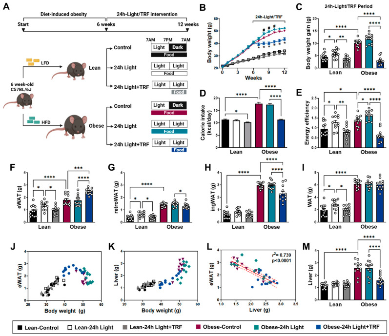
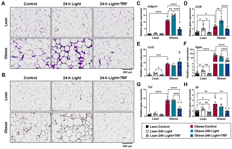
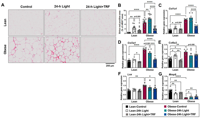
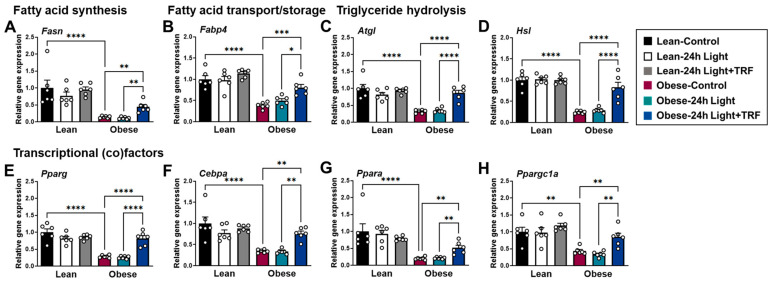
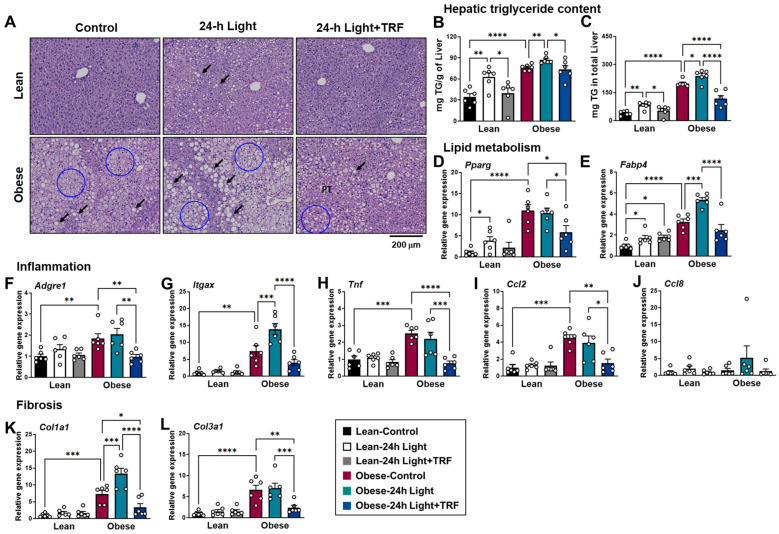
Time-restricted feeding (TRF) has emerged as a promising dietary approach for improving metabolic parameters associated with obesity. However, it remains largely unclear whether TRF offers benefits for obesity related to exposure to light at night. This study examined whether lean and obese mice under chronic light exposure could benefit from TRF intervention. Six-week-old C57BL/6 male mice were fed either a low-fat diet or a high-fat diet under a 12 h light/12 h dark cycle for 6 weeks. They were then divided into three subgroups: control light, chronic 24 h light, and chronic light with a daily 10 h TRF. Chronic light exposure led to increased weight gain and higher expression of inflammatory and fibrotic markers in the adipose tissue of both lean and obese mice. It also increased hepatic triglyceride content in mice, regardless of their weight status. TRF protected both lean and obese mice from weight gain, normalized inflammatory and fibrotic gene expression, and reduced adipose tissue collagen and liver triglyceride accumulation caused by light exposure alone or in combination with obesity. These results suggest that TRF could have clinical implications for preventing obesity associated with night shift work, regardless of current weight status.

Keywords: adipose tissue inflammation; chronic light; fibrosis; high-fat diet; time-restricted feeding.


Conflict of interest statement

The authors declare no conflicts of interest.

Figures

Figure 1
Figure 1
Effects of chronic light exposure and time-restricted feeding (TRF) on body weight and tissue mass. (A) Overview of the study design. Six-week-old C57BL/6J male mice were fed either a low-fat diet (LFD) or a high-fat diet (HFD) ad libitum under a control light condition (12 h light/dark cycle). After 6 weeks, the LFD-fed lean or HFD-fed obese mice were exposed for another 6 weeks to either a control light condition (12 h light/dark cycle), chronic 24 h light exposure, or chronic 24 h light exposure with a daily 10 h TRF intervention. (B) Body weight during the entire period. # p < 0.05 for Obese-24 h Light (vs. Obese-control); * p < 0.05 for Obese-24 h Light+TRF (vs. Obese-24 h Light). (C) Body weight gain, (D) daily calorie intake, and (E) energy efficiency ratio during the 6-week chronic light exposure and TRF intervention period. (FI) Adipose tissue weight: (F) epididymal white adipose tissue (eWAT), (G) retroperitoneal white adipose tissue (retroWAT), (H) inguinal white adipose tissue (ingWAT), and (I) total adipose tissue (the sum of eWAT, retroWAT, and ingWAT). (J) eWAT weight and (K) liver mass are expressed as a function of body weight. (L) Inverse association between eWAT mass and liver weight in obese mice. (M) Liver weight. Lean-control: Mice fed a LFD ad libitum for 12 weeks under a control light condition (12 h light/dark cycle); Lean-24 h Light: Mice fed a LFD ad libitum for 12 weeks but exposed to chronic 24 h light for the last 6 weeks; Lean-24 h Light+TRF: Mice fed a LFD ad libitum for 6 weeks, then exposed to chronic 24 h light and a daily 10 h TRF for the last 6 weeks; Obese-control: Mice fed a HFD ad libitum for 12 weeks under a control light condition (12 h light/dark cycle); Obese-24 h Light: Mice fed a HFD ad libitum for 12 weeks but exposed to chronic 24 h light for the last 6 weeks; Obese-24 h Light+TRF: Mice fed a HFD ad libitum for 6 weeks, then exposed to chronic 24 h light and a daily 10 h TRF for the last 6 weeks. Data are presented as means ± SEM, n = 12 mice per group. * p < 0.05, ** p < 0.01, *** p < 0.001, **** p < 0.0001 by ANOVA with Tukey’s post hoc test.
Figure 2
Figure 2
Effects of chronic light exposure and TRF on adipose tissue inflammation. (A,B) Representative images of epididymal adipose tissue stained with (A) H&E and (B) anti-F4/80 antibody. Scale bar = 200 μm. (CH) Quantitative real-time PCR analysis of (C) macrophage-specific marker gene (Adgre1), (D) monocyte chemoattractant protein 2 (Ccl8), (E) monocyte chemoattractant protein 1 (Ccl2), (F) proinflammatory macrophage marker gene (Itgax), (G) proinflammatory cytokine (Tnf), and (H) proinflammatory cytokine (Il6) in epididymal fat. Data are presented as means ± SEM, n = 6 mice per group. * p < 0.05, ** p < 0.01, *** p < 0.001, **** p < 0.0001 by ANOVA with Tukey’s post hoc test.
Figure 3
Figure 3
Effects of chronic light exposure and TRF on adipose tissue fibrosis. (A) Representative images of Picrosirius red-stained collagen in epididymal adipose tissues. Scale bar = 200 μm. (B) Picrosirius red-positive area. Data are presented as means ± SEM, n = 12 mice per group. (CG) Quantitative real-time PCR analysis of (C) Col1a1, (D) Col3a1, (E) Col6a3, (F) Lox, and (G) Mmp9 in epididymal fat. Data are presented as means ± SEM, n = 6 mice per group. * p < 0.05, ** p < 0.01, *** p < 0.001, **** p < 0.0001 by ANOVA with Tukey’s post hoc test.
Figure 4
Figure 4
Effects of chronic light exposure and TRF on the expression of genes involved in lipid metabolism in epididymal adipose tissue. Quantitative real-time PCR analysis of (A) fatty acid synthase (Fasn), (B) fatty acid-binding protein 4 (Fabp4), (C) adipose triglyceride lipase (Atgl), (D) hormone-sensitive lipase (Hsl), and (EH) transcriptional (co)factors in epididymal fat. Data are presented as means ± SEM, n = 6 mice per group. * p < 0.05, ** p < 0.01, *** p < 0.001, **** p < 0.0001 by ANOVA with Tukey’s post hoc test.
Figure 5
Figure 5
Effects of chronic light exposure and TRF on hepatic steatosis, inflammation, and fibrosis. (A) Representative H&E-stained histological sections of liver tissues. Macrovesicular lipid droplets (arrows) and microvesicular steatosis (circle) are present during the development of liver steatosis. Scale bar = 200 μm. (B,C) Hepatic triglyceride content adjusted by liver weight (B) or in total liver weight (C). (DL) Quantitative real-time PCR analysis of (D) Pparg, (E) Fabp4, (F) Adgre1, (G) Itgax, (H) Tnf, (I) Ccl2, (J) Ccl8, (K) Col1a1, and (L) Col3a1 in liver. Data are presented as means ± SEM, n = 6 mice per group. * p < 0.05, ** p < 0.01, *** p < 0.001, **** p < 0.0001 by ANOVA with Tukey’s post hoc test.

Similar articles

Cited by

  • The diverse roles of the circadian clock in cancer.
    Fortin BM, Mahieu AL, Fellows RC, Kang Y, Lewis AN, Ead AS, Lamia KA, Cao Y, Pannunzio NR, Masri S. Fortin BM, et al. Nat Cancer. 2025 May;6(5):753-767. doi: 10.1038/s43018-025-00981-8. Epub 2025 May 26. Nat Cancer. 2025. PMID: 40419761 Free PMC article. Review.

References

    1. Muscogiuri G., Poggiogalle E., Barrea L., Tarsitano M.G., Garifalos F., Liccardi A., Pugliese G., Savastano S., Colao A., Alviggi C., et al. Exposure to artificial light at night: A common link for obesity and cancer? Eur. J. Cancer. 2022;173:263–275. doi: 10.1016/j.ejca.2022.06.007. - DOI - PubMed
    1. Rybnikova N.A., Haim A., Portnov B.A. Does artificial light-at-night exposure contribute to the worldwide obesity pandemic? Int. J. Obes. 2016;40:815–823. doi: 10.1038/ijo.2015.255. - DOI - PubMed
    1. Fonken L.K., Workman J.L., Walton J.C., Weil Z.M., Morris J.S., Haim A., Nelson R.J. Light at night increases body mass by shifting the time of food intake. Proc. Natl. Acad. Sci. USA. 2010;107:18664–18669. doi: 10.1073/pnas.1008734107. - DOI - PMC - PubMed
    1. Park Y.-M.M., White A.J., Jackson C.L., Weinberg C.R., Sandler D.P. Association of Exposure to Artificial Light at Night While Sleeping with Risk of Obesity in Women. JAMA Intern. Med. 2019;179:1061–1071. doi: 10.1001/jamainternmed.2019.0571. - DOI - PMC - PubMed
    1. Brum M.C.B., Filho F.F.D., Schnorr C.C., Bertoletti O.A., Bottega G.B., Rodrigues T.d.C. Night shift work, short sleep and obesity. Diabetol. Metab. Syndr. 2020;12:1–9. doi: 10.1186/s13098-020-0524-9. - DOI - PMC - PubMed

LinkOut - more resources