Comparative Study
. 2024 Oct 26;25(21):11526.
doi: 10.3390/ijms252111526.

Comparative Analysis of Commercial and Home-Made Media on RSPO1/S6R Axis in Organoids with Different Wnt Backgrounds: A Methodological Guide for the Selection of Intestinal Patient-Derived Organoids Culture Media

Affiliations
Comparative Study

Comparative Analysis of Commercial and Home-Made Media on RSPO1/S6R Axis in Organoids with Different Wnt Backgrounds: A Methodological Guide for the Selection of Intestinal Patient-Derived Organoids Culture Media

Giulia Calafato et al. Int J Mol Sci. .

Abstract

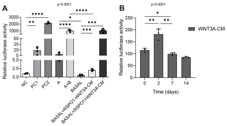
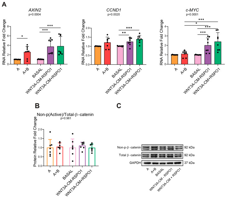
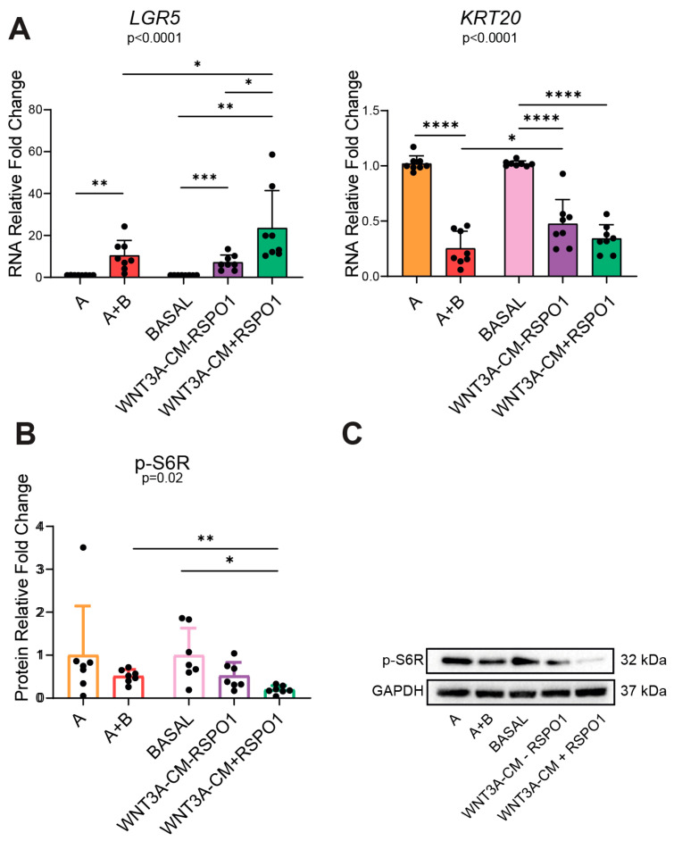
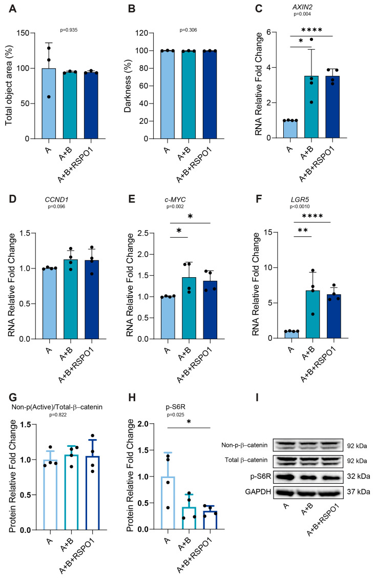
WNT3A is an intestinal ligand triggering the Wnt/β-catenin (Wnt) pathway, which can be enhanced by R-spondin 1 (RSPO1) through the RSPO1-LGR axis or antagonized by the adenomatous polyposis coli (APC) protein supporting β-catenin-degradation. Wnt interplays with several pathways including PI3K/mTOR (mTOR). In this study, we evaluated the influence of WNT3A-commercial and home-made culture media and RSPO1 protein on the Wnt and mTOR interplay in non-APC and APC-mutated intestinal patient-derived organoids (PDOs). Normal mucosa (NM) of sporadic CRC and FAP PDOs were cultured with: WNT3A-lacking/containing commercial (A/A+B) or home-made (BASAL/WNT3A-conditioned medium (CM)±RSPO1) media. In non-APC-mutated-PDOs (CRC-NM), WNT3A-CM, over commercial A+B, strongly activated Wnt-target-genes CCND1 and c-MYC. Most importantly, the addition of RSPO1 to home-made WNT3A-CM or A+B led to the downregulation of the mTOR-downstream-effector phospho-S6 ribosomal protein (p-S6R), highlighting the activation of the RSPO1-pS6R in both non-APC (CRC-NM) and APC-mutated (FAP-NM) PDOs, independently from LGR5 gene expression modulation. Our work demonstrates that home-made WNT3A-CM strongly impacts the crosstalk between Wnt and mTOR over commercial media, and proposes RSPO1 as a key regulator of the RSPO1-p-S6R axis in both non-APC and APC-mutated PDOs. Together, these findings represent an important methodological guide for scientists working in these fields to select the most appropriate intestinal PDO media.

Keywords: R-spondin 1 (RSPO1); colorectal cancer (CRC); intestinal patient-derived organoid (PDO); organoids culture media; phospho-S6 ribosomal protein (p-S6R).


Conflict of interest statement

The authors declare no conflicts of interest. The funders had no role in the study design, data collection and analysis, decision to publish, or preparation of the manuscript.

Figures

Figure 1
Figure 1
Canonical Wnt/β-catenin pathway activation in the HEK 293 STF cell reporter cell line in commercial and home-made-cultured non-APC mutated CRC NM PDOs. (A) Luciferase assay of HEK 293 STF cells cultured for 24 h with different intestinal organoid culture media (n = 4). (B) Luciferase-based quality control test of WNT3A-CM in HEK 293 STF cells cultured for 24 h with freshly thawed medium (termed 0) or 2/7/14-days-thawed medium (n = 3). Experiments were independently repeated at least three times and the values are reported as the mean ± SD. Data were analyzed with the parametric Student’s t-test according to Shapiro–Wilk normality test results. * p < 0.05, ** p  < 0.01, *** p  < 0.001, **** p < 0.0001. NC = negative control; PC1 = positive control 1; PC2 = positive control 2; A = WNT3A-lacking commercial medium composed of DMEMF12 and Component A in a 1:1 ratio; A+B = WNT3A-containing commercial medium composed of Component A and Supplement B in a 1:1 ratio (exact formulation relative to other factors present is not known); BASAL = WNT3A-lacking home-made basal medium; WNT3A-CM = WNT3A-containing complete home-made medium, with or without R-spondin1 (+/−RSPO1).
Figure 2
Figure 2
Different activation of Wnt/β-catenin in commercial and home-made-cultured non-APC mutated CRC NM PDOs. (A) RNA expression of Wnt/β-catenin target genes AXIN2, CCND1, and c-MYC of CRC NM PDOs cultured for 48 h with the indicated media (n = 8, of which PT10 = 6, PT13 = 1, PT16 = 1). (B) WB analysis of non-p(Active)/total β-catenin protein ratio in CRC NM PDOs cultured for 48 h with the specified media as a marker of Wnt activation (n = 7, of which PT10 = 5, PT13 = 1, PT16 = 1). (C) Representative WB imaging of non-p(Active)-β-catenin, total β-catenin, p-S6R, and GAPDH (n = 7); GAPDH was used as the loading control (cropped blots). Experiments were independently repeated at least three times and the values are reported as mean ± SD. Data were analyzed with the parametric Student’s t-test (CCND1 and non-p(Active)/total β-catenin) and non-parametric Mann–Whitney test (AXIN2 and c-MYC), according to the Shapiro–Wilk normality test results. * p < 0.05, ** p  < 0.01 and *** p  < 0.001. A = WNT3A-lacking commercial medium composed of DMEMF12 and Component A in a 1:1 ratio; A+B = WNT3A-containing commercial medium composed of Component A and Supplement B in a 1:1 ratio (exact formulation relative to other factors present is not known); BASAL = WNT3A-lacking home-made basal medium; WNT3A-CM = WNT3A-containing complete home-made medium, with or without R-spondin1 (+/−RSPO1).
Figure 3
Figure 3
Different activation of intestinal stemness/differentiation markers and PI3K/mTOR in commercial and home-made-cultured non-APC mutated CRC NM PDOs. (A) RNA expression of stemness (LGR5) and differentiation (KRT20) intestinal markers in 48 h-cultured CRC NM PDOs (n = 8, of which PT10 = 6, PT13 = 1, PT16 = 1). (B) WB analysis of mTOR effector p-S6R protein in CRC NM PDOs cultured for 48 h with the indicated media (n = 7, of which PT10 = 5, PT13 = 1, PT16 = 1). (C) Representative WB imaging of non-p(Active)-β-catenin, total β-catenin, p-S6R, and GAPDH (n = 7); GAPDH was used as the loading control (cropped blots). Experiments were independently repeated at least three times and the values are reported as mean ± SD. Data were analyzed with the parametric Student’s t-test (LGR5 and KRT20) and non-parametric Mann–Whitney test (p-S6R), according to the Shapiro–Wilk normality test results. * p < 0.05, ** p  < 0.01, *** p  < 0.001, **** p < 0.0001. A = WNT3A-lacking commercial medium composed of DMEMF12 and Component A in a 1:1 ratio; A+B = WNT3A-containing commercial medium composed of Component A and Supplement B in a 1:1 ratio (exact formulation relative to other factors present is not known); BASAL = WNT3A-lacking home-made basal medium; WNT3A-CM = WNT3A-containing complete home-made medium, with or without R-spondin1 (+/−RSPO1).
Figure 4
Figure 4
Effect of commercial and home-made media on the viability and morphology of non-APC-mutated CRC NM PDOs. (A) Representative images of CRC NM PDOs cultured with the indicated media at 48 h (40× magnification, scale bar = 100 µm) (n = 6); representative pictures were captured through an EVOS M5000 Microscope. (B) Analysis of CRC NM PDO viability cultured for 48 h with the different culture media (n = 6, of which PT10 = 4, PT13 = 1, PT16 = 1). (C) Morphological analysis of the total object area and darkness of CRC NM PDOs in different culture media at the 48 h time point (n = 6, of which PT10 = 4, PT13 = 1, PT16 = 1). (D) Time-lapse morphological analysis of cultured-CRC NM PDOs. Morphology (total object area and darkness) was monitored every 8 h for 48 h (n = 6, of which PT10 = 4, PT13 = 1, PT16 = 1). (E,F) WB analysis and representative imaging of SURVIVIN protein in CRC NM PDOs cultured for 48 h with the specified media (n = 6, of which PT10 = 4, PT13 = 1, PT16 = 1); GAPDH was used as the loading control (cropped blots). Experiments were independently repeated six times and values are shown as the mean ± SD. Data were analyzed with the parametric Student’s t-test (viability assay and morphological analysis) and non-parametric Mann–Whitney test (SURVIVIN), according to the Shapiro–Wilk normality test results. A = WNT3A-lacking commercial medium composed of DMEMF12 and Component A in a 1:1 ratio; A+B = WNT3A-containing commercial medium composed of Component A and Supplement B in a 1:1 ratio (exact formulation relative to other factors present is not known); BASAL = WNT3A-lacking home-made basal medium; WNT3A-CM = WNT3A-containing complete home-made medium, with or without R-spondin1 (+/−RSPO1).
Figure 5
Figure 5
A+B-cultured-APC-mutated FAP NM PDOs induce lower LGR5 activation without affecting p-S6R downregulation. (A) RNA expression analysis of the Wnt/β-catenin target genes AXIN2, CCND1, and c-MYC in CRC NM and FAP NM PDOs cultured for 48 h in A and A+B (n = 5 of which, FAP NM-01 = 2, FAP NM-03 = 3, PT10 CRC NM = 3, PT13 CRC NM = 1, PT16 CRC NM = 1). (B) RNA expression analysis of stemness (LGR5) and differentiation (KRT20) intestinal markers in CRC NM and FAP NM PDOs cultured for 48 h in A and A+B (n = 5 of which, FAP NM-01 = 2, FAP NM-03 = 3, PT10 CRC NM = 3, PT13 CRC NM = 1, PT16 CRC NM = 1). (C,D) WB analysis of non-p(Active)/total β-catenin protein ratio and p-S6R in CRC NM and FAP NM PDOs cultured for 48 h with A and A+B (n = 5 of which, FAP NM-01 = 2, FAP NM-03 = 3, PT10 CRC NM = 3, PT13 CRC NM = 1, PT16 CRC NM = 1). (E) Representative WB imaging of non-p(Active)-β-catenin, total β-catenin, p-S6R, and GAPDH in CRC NM and FAP NM PDOs cultured for 48 h with A and A+B (n = 5 of which, FAP NM-01 = 2, FAP NM-03 = 3, PT10 CRC NM = 3, PT13 CRC NM = 1, PT16 CRC NM = 1); Representative WB imaging of FAP NM (A/A+B) and CRC NM (A/A+B) (cropped blots). GAPDH was used as the loading control. Experiments were independently repeated five times and values are shown as mean ± SD. Data were analyzed with the parametric Student’s t-test (AXIN2, c-MYC, CCND1, LGR5, non-p(Active)/total β-catenin and p-S6R) and non-parametric Mann–Whitney test (KRT20), according to the Shapiro–Wilk normality test results. * p < 0.05, ** p  < 0.01. A = WNT3A-lacking commercial medium composed of DMEMF12 and Component A in a 1:1 ratio; A+B = WNT3A-containing commercial medium composed of Component A and Supplement B in a 1:1 ratio (exact formulation relative to other factors present is not known).
Figure 6
Figure 6
RSPO1 guides p-S6R downregulation in APC-mutated FAP NM PDOs independently from a boosted activation of LGR5. (A,B) Morphological analysis of the total object area and darkness of FAP NM PDOs cultured with the indicated media for 48 h (n = 3, FAP NM-03 = 3). (CF) RNA expression analysis of the AXIN2, CCND1, c-MYC Wnt-target genes and LGR5 intestinal marker in FAP NM PDOs cultured for 48 h with A, A+B, and A+B+RSPO1 (n = 4, FAP NM-03 = 4). (G,H) WB analysis of non-p(Active)/total β-catenin protein ratio and p-S6R in FAP NM PDOs cultured for 48 h with the indicated media (n = 4, FAP NM-03 = 4). (I) Representative WB imaging of non-p(Active)-β-catenin, total β-catenin, p-S6R, and GAPDH in FAP NM PDOs cultured for 48 h with the indicated media (n = 4, FAP NM-03 = 4); GAPDH was used as the loading control (cropped blots). Experiments were independently repeated at least three times and values are shown as the mean ± SD. Data were analyzed with the parametric Student’s t-test (morphological analysis, LGR5, non-p(Active)/total β-catenin and p-S6R), according to the Shapiro–Wilk normality test results. * p < 0.05, ** p  < 0.01, **** p < 0.0001. A = WNT3A-lacking commercial medium composed of DMEMF12 and Component A in a 1:1 ratio; A+B = WNT3A-containing commercial medium composed of Component A and Supplement B in a 1:1 ratio (exact formulation relative to other factors present is not known); A+B+RSPO1 = WNT3A-containing commercial medium composed of Component A and Supplement B in a 1:1 ratio, with RSPO1 3.

Similar articles

References

    1. Liu J., Xiao Q., Xiao J., Niu C., Li Y., Zhang X., Zhou Z., Shu G., Yin G. Wnt/β-Catenin Signalling: Function, Biological Mechanisms, and Therapeutic Opportunities. Signal Transduct. Target. Ther. 2022;7:3. doi: 10.1038/s41392-021-00762-6. - DOI - PMC - PubMed
    1. De Lau W., Barker N., Low T.Y., Koo B.K., Li V.S.W., Teunissen H., Kujala P., Haegebarth A., Peters P.J., Van De Wetering M., et al. Lgr5 Homologues Associate with Wnt Receptors and Mediate R-Spondin Signalling. Nature. 2011;476:293–297. doi: 10.1038/nature10337. - DOI - PubMed
    1. Clevers H. Wnt/Beta-Catenin Signaling in Development and Disease. Cell. 2006;127:469–480. doi: 10.1016/j.cell.2006.10.018. - DOI - PubMed
    1. Fodde R., Smits R., Clevers H. APC, Signal Transduction and Genetic Instability in Colorectal Cancer. Nat. Rev. Cancer. 2001;1:55–67. doi: 10.1038/35094067. - DOI - PubMed
    1. McGough I.J., Vincent J.P. APC Moonlights to Prevent Wnt Signalosome Assembly. Dev. Cell. 2018;44:535–537. doi: 10.1016/j.devcel.2018.02.018. - DOI - PMC - PubMed

Publication types

MeSH terms

LinkOut - more resources