. 2024 Oct 27;25(21):11538.
doi: 10.3390/ijms252111538.

Cartilage Laser Engraving for Fast-Track Tissue Engineering of Auricular Grafts

Affiliations

Cartilage Laser Engraving for Fast-Track Tissue Engineering of Auricular Grafts

Anastas A Kisel et al. Int J Mol Sci. .

Abstract

In this study, the optimal engraving parameters were determined through the analysis of scanning electron microscopy (SEM) data, as follows: a laser power density of 5.5 × 10⁵ W/cm2, an irradiation rate of 0.1 mm/s, a well radius of 60 μm, a distance between well centers of 200 μm, and a number of passes for each well of 20. After 1 week of in vitro cultivation, chondrocytes were located on the surface of the scaffolds, in the sockets and lacunae of decellularized cartilage. When implanted into animals, both cellular and acellular scaffolds were able to support cartilage in-growth and complete regeneration of the defect without clear boundaries with normal tissue. Nevertheless, the scaffolds populated with cells exhibited superior biocompatibility and were not subject to rejection, in contrast to cell-free constructs.

Keywords: ear cartilage; laser engraving; nasal chondrocytes; regenerative medicine; tissue engineering.


Conflict of interest statement

The authors declare no conflict of interest.

Figures

Figure 1
Figure 1
Laser engraving of the devitalized ear cartilage. (I) The principle of laser engraving of the devitalized ear cartilage. (II) Probability density of perforation depth, n = 132. The red line shows the probability density curve; (III) Light microscopy of dECM laser engraving, objective lens ×5, scale bar 250 μm. (IV) SEM laser-engraved dECM of rabbit auricle, scale bar 300 μm. (V) SEM laser-engraved dECM of rabbit auricle, scale bar 100 μm.
Figure 2
Figure 2
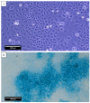
Chondrocytes of the 0th passage after 7 days of cultivation in a Petri dish. (I) Phase contrast, objective lens ×10, scale bar—200 μm. (II) Alcian blue staining, glycosaminoglycans. Objective lens ×20, scale bar—100 μm.
Figure 3
Figure 3
A cross-section of LPECs populated with nasal chondrocytes after 7 days of in vitro cultivation. Hematoxylin and eosin staining. (I) The black arrows indicate a formed non-square perforation, objective lens ×10, scale bar—200 μm. (II) The white arrows indicate the layer of attached nasal chondrocytes, objective lens ×20, scale bar—100 μm. (III) The white arrows show cells filling the perforation in LPECs, objective lens ×40, scale bar—50 μm. A longitudinal section of LPECs populated with nasal chondrocytes after 7 days of in vitro cultivation. Safranin-O staining. (IV) The white arrows indicate the formation of the nasal chondrocyte layer, objective lens ×10, scale bar—200 μm. (V) GAG production in the intercellular substance of repopulated chondrocytes (asterisk), objective lens ×20, scale bar—100 μm. (VI) Objective lens ×40, scale bar—50 μm.
Figure 4
Figure 4
Cell-free LPECs. (I) A longitudinal section of LPECs, objective lens ×10, scale bar—200 μm. Hematoxylin and eosin staining. (II) Objective lens ×20, scale bar—100 μm. Safranin-O staining. (III) Objective lens ×10, scale bar—200 μm. Safranin-O staining. (IV) Objective lens ×20, scale bar—100 μm. Safranin-O staining.
Figure 5
Figure 5
In vivo orthotopic study. LPEC transplant surgery. General view of the operated area. Creation of a cartilage defect (I), placement of LPEC in the defect (II) and the suturing of the operated area (III); general view of the surgical wound with nasal chondrocyte cells populated (IV) and control group (V) in rabbit auricles. Rejected graft of the control group in the rabbit’s auricles (VI).
Figure 6
Figure 6
Transverse section of cartilage tissue of the control (I,II) and experimental (III,IV) groups. (A) Hematoxylin and eosin staining, (B) Safranin-O staining and (C) Immunohistochemical staining for type II collagen. Objective lens ×10, scale bar—200 μm (I,III) and objective lens ×20, scale bar—100 μm (II,IV).
Figure 7
Figure 7
(A) An overgrowth of newly formed cartilage tissue. The dotted area indicates LPECs. White arrows indicate single chondroblasts. Hematoxylin and eosin staining. Objective lens ×40, scale bar—50 μm; (B) the inflammatory responses and resorption of LPECs. (I) Inflammatory infiltrates in the area of rejection LPECs. Cellular infiltrate (asterisk). Hematoxylin and eosin staining, objective lens ×10, scale bar—200 μm. (II) Resorption of implanted LPECs. The arrow indicates the remains of the structure. Hematoxylin and eosin staining, objective lens ×10, scale bar—200 μm and (III) Resorption of implanted LPECs. The arrow indicates the remains of the structure. Hematoxylin and eosin staining, objective lens ×20, scale bar—100 μm.

References

    1. Sobol E., Shekhter A., Guller A., Baum O., Baskov A. Laser-induced regeneration of cartilage. J. Biomed. Opt. 2011;16:080902. doi: 10.1117/1.3614565. - DOI - PubMed
    1. Humphries S., Joshi A., Webb W.R., Kanegaonkar R. Auricular reconstruction: Where are we now? A critical literature review. Eur. Arch. Oto-Rhino-Laryngol. 2022;279:541–556. doi: 10.1007/s00405-021-06903-5. - DOI - PubMed
    1. Beketov E.E., Isaeva E.V., Yakovleva N.D., Demyashkin G.A., Arguchinskaya N.V., Kisel A.A., Lagoda T.S., Malakhov E.P., Kharlov V.I., Osidak E.O., et al. Bioprinting of Cartilage with Bioink Based on High-Concentration Collagen and Chondrocytes. Int. J. Mol. Sci. 2021;22:11351. doi: 10.3390/ijms222111351. - DOI - PMC - PubMed
    1. Pina S., Ribeiro V.P., Marques C.F., Maia F.R., Silva T.H., Reis R.L., Oliveira J.M. Scaffolding strategies for tissue engineering and regenerative medicine applications. Materials. 2019;12:1824. doi: 10.3390/ma12111824. - DOI - PMC - PubMed
    1. Ovsianikov A., Khademhosseini A., Mironov V. The Synergy of Scaffold-Based and Scaffold-Free Tissue Engineering Strategies. Trends Biotechnol. 2018;36:348–357. doi: 10.1016/j.tibtech.2018.01.005. - DOI - PubMed

LinkOut - more resources