. 2024 Oct 30;25(21):11667.
doi: 10.3390/ijms252111667.

Chitin Translocation Is Functionally Coupled with Synthesis in Chitin Synthase

Affiliations

Chitin Translocation Is Functionally Coupled with Synthesis in Chitin Synthase

Suhao Niu et al. Int J Mol Sci. .

Abstract

Chitin, an extracellular polysaccharide, is synthesized by membrane-embedded chitin synthase (CHS) utilizing intracellular substrates. The mechanism of the translocation of synthesized chitin across the membrane to extracellular locations remains unresolved. We prove that the chitin synthase from Phytophthora sojae (PsCHS) is a processive glycosyltransferase, which can rapidly produce and tightly bind with the highly polymerized chitin. We further demonstrate that PsCHS is a bifunctional enzyme, which is necessary and sufficient to translocate the synthesized chitin. PsCHS was purified and then reconstituted into proteoliposomes (PLs). The nascent chitin is generated and protected from chitinase degradation unless detergent solubilizes the PLs, showing that PsCHS translocates the newly produced chitin into the lumen of the PLs. We also attempted to resolve the PsCHS structure of the synthesized chitin-bound state, although it was not successful; the obtained high-resolution structure of the UDP/Mn2+-bound state could still assist in describing the characterization of the PsCHS's transmembrane channel. Consistently, we demonstrate that PsCHS is indispensable and capable of translocating chitin in a process that is tightly coupled to chitin synthesis.

Keywords: chitin synthase; glycosyltransferase; membrane translocation; processive.


Conflict of interest statement

The authors declare no conflicts of interest.

Figures

Figure 1
Figure 1
Processivity analysis of PsCHS. (A) The size exclusion chromatography profile and SDS–PAGE analysis of purified PsCHS is displayed, with peak position indicated by the asterisk and purified PsCHS marked by the red arrow. (B) The purified PsCHS shows activity in vitro (Column 1). Chitinase was added after the synthesis reaction, proving that the synthesized product is chitin (Column 2). Incubation with EDTA can inactivate PsCHS (Column 3). (C) The processive property of PsCHS was verified using a sedimentation experiment assay. Western blot analysis indicates that PsCHS is present in the precipitate (P) after the chitin synthesis reaction (Lane 4). PsCHS does not self-precipitate (Lane 2) or interact with the exogenous chitin (Lane 6) and only exists in the supernatant (S) in the control experiments (Lanes 1 and 5). S and P refer to the supernatant and precipitate of centrifugation, respectively. (D) The pulse-chase analysis of PsCHS. The pulse samples were extracted from a reaction mixture with a low enzyme-to-substrate molar ratio of 1:3 at a specific time. Then, the enzyme-to-substrate molar ratio was increased to 1:1000 by supplementing the reaction mixture with additional substrate and continuing incubation. The chase samples were removed at the indicated time. The distribution of products was analyzed by scintillation counting after filter paper chromatography. DPM, disintegrations per minute. (E) Scheme illustrating the distribution of product lengths for processive and distributive glycosyltransferases. The monosaccharide units are depicted as hexagons, with labeled sugars in brown and unlabeled sugars in orange.
Figure 2
Figure 2
Chitin translocation assay using PsCHS. (A) Schematic representation of the in vitro chitin translocation assay. Chitin is only synthesized when the GT domain is outward facing but not inward facing. Translocation of chitin across the vesicle membrane separates the polymer from the solvent, which can be quantified by the enzymatic degradation of chitin. Without translocation, chitin will be susceptible to digestion by chitinase addition, whereas translocated chitin will only be digested after solubilizing the lipid vesicles with detergent. (B) Western blot of proteoliposomes (PLs) embedding PsCHS, with positive bands of PsCHS indicated by the red arrow. Western blot analysis indicates that PsCHS co-precipitates with PLs after ultracentrifugation, demonstrating that purified PsCHS was successfully reconstituted into E. coli total lipid extract PLs. PLs, PsCHS embedded proteoliposomes; PLs-S, supernatant part after PsCHS embedded proteoliposomes ultracentrifugation; PLs-P, precipitate part after PsCHS embedded proteoliposomes ultracentrifugation. (C) In vitro chitin translocation function of PsCHS. PLs containing PsCHS were incubated with a radioactively labeled substrate, and then, the reaction was terminated with EDTA. Subsequently, the PLs were incubated with chitinase in the presence or absence of Triton X-100 followed by inactivation of chitinase with SDS. No chitin is synthesized when EDTA is added to the reaction mixture before PsCHS addition (column 4), confirming that EDTA efficiently terminates chitin elongation. Chitin is quantified relative to the amount obtained without enzymatic digestion. (D) Degradation of chitin within the detergent environment. Similar experiments to panel C were conducted, except that PsCHS in detergent was used instead of PsCHS embedded in PLs. Chitin was completely digested in either the presence or absence of Triton X-100.
Figure 3
Figure 3
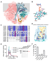
The structure and chitin translocation channel of PsCHS. (A) The architecture of the PsCHS. The side view of the PsCHS dimer is shown in surface and cartoon representations. The TM helices, IF helices, GT domain, and N-terminal domain associated with dimerization are colored as pale blue, indigo blue, dark turquoise, and golden yellow, respectively. The other protomer is colored as pink. The unresolved region is shown as dashed lines. The approximate position of the membrane is marked with grey shading. (B) Interactions between PsCHS and UDP/Mn2+. UDP is stabilized in the reaction cavity of the GT domain through hydrogen bonds and π-π stacking hydrophobic interaction. Mn2⁺ coordinates with the pyrophosphate group of UDP. The interactions between PsCHS and UDP/Mn2+ were analyzed using Protein–Ligand Interaction Profiler (PLIP) [21]. Residues are colored according to their respective functional regions, consistent with Figure 3A. Hydrogen bonds, π-π stacking interactions, and coordinates between PsCHS and UDP/Mn2+ are indicated with blue lines, green dashed lines, and orange lines, respectively. (C) Sequence alignment of PsCHS with chitin synthases from other species shows that three residues potentially involved in chitin translocation in PsCHS are conserved among chitin synthases from Saccharomyces cerevisiae (Sc), Peronospora effusa (Pe), Candida albicans (Ca), Sporothrix brasiliensis (Sb), and Coccidioides immitis (Ci). These conserved residues are marked with red squares and highlighted in panel D. (D) The chitin translocation channel of PsCHS. The presumed chitin translocation channel is illustrated with a golden surface, and the selected conserved amino acids at different positions are shown by sticks. The colors match those in panel A, with the selected residues highlighted in red. (E) Purification of PsCHS mutations. The size exclusion chromatography profile for PsCHS and its mutants was obtained using a Superdex 200 Increase 10/300 GL column. The lines in red, blue, green, and black represent the mutants L540A, Y698A, V832A, and wild-type PsCHS, respectively, with peak position indicated by the asterisk. The positive bands in Western blot of purified PsCHS and its mutants is indicated by the red arrow. (F) The effect of mutations on PsCHS. Mutating the selected amino acids significantly reduced the activity of chitin synthase.

Similar articles

References

    1. Brown H.E., Esher S.K., Alspaugh J.A. Chitin: A “Hidden Figure” in the fungal cell wall. Curr. Top. Microbiol. Immunol. 2020;425:83–111. - PubMed
    1. Merzendorfer H. Insect chitin synthases: A review. J. Comp. Physiol. B. 2005;176:1–15. doi: 10.1007/s00360-005-0005-3. - DOI - PubMed
    1. Drula E., Garron M.-L., Dogan S., Lombard V., Henrissat B., Terrapon N. The carbohydrate-active enzyme database: Functions and literature. Nucleic Acids Res. 2022;50:D571–D577. doi: 10.1093/nar/gkab1045. - DOI - PMC - PubMed
    1. Coutinho P.M., Deleury E., Davies G.J., Henrissat B. An evolving hierarchical family classification for glycosyltransferases. J. Mol. Biol. 2003;328:307–317. doi: 10.1016/S0022-2836(03)00307-3. - DOI - PubMed
    1. Campbell J.A., Davies G.J., Bulone V., Henrissat B. A classification of nucleotide-diphospho-sugar glycosyltransferases based on amino acid sequence similarities. Biochem. J. 1997;326:929–939. doi: 10.1042/bj3260929u. - DOI - PMC - PubMed

LinkOut - more resources