. 2024 Oct 31;25(21):11709.
doi: 10.3390/ijms252111709.

Kinin Receptors B1 and B2 Mediate Breast Cancer Cell Migration and Invasion by Activating the FAK-Src Axis

Affiliations

Kinin Receptors B1 and B2 Mediate Breast Cancer Cell Migration and Invasion by Activating the FAK-Src Axis

Felipe González-Turén et al. Int J Mol Sci. .

Abstract

Kinin receptors B1 and B2 are involved in migration and invasion in gastric, glioma, and cervical cancer cells, among others. However, the role of kinin receptors in breast cancer cells has been poorly studied. We aimed to reveal the impact of B1 and B2 receptors on migration and invasion in breast cancer cells and demonstrate their capacity to modulate in vivo tumor growth. MDA-MB-231, MCF-7, and T47D cells treated with Lys-des[Arg9]bradykinin (LDBK) or bradykinin (BK) were used to evaluate migration and invasion. Des-[Arg9]-Leu8-BK and HOE-140 were used as antagonists for the B1 and B2 receptors. MDA-MB-231 cells incubated or not with antagonists were subcutaneously inoculated in BALBc NOD/SCID mice to evaluate tumor growth. LDBK and BK treatment significantly increased migration and invasion in breast cancer cells, effects that were negated when antagonists were used. The use of antagonists in vivo inhibited tumor growth. Moreover, the migration and invasion induced by kinins in breast cancer cells were inhibited when focal adhesion kinase (FAK) and Src inhibitors were used. The novelty revealed in our work is that B1 and B2 receptors activated by LDBK and BK induce migration and invasion in breast cancer cells via a mechanism that involves the FAK-Src signaling pathway, and the antagonism of both receptors in vivo impairs breast tumor growth.

Keywords: B1 receptor; B2 receptor; FAK; Src; breast cancer; invasion; kinin receptors; migration.


Conflict of interest statement

The authors declare no conflicts of interest.

Figures

Figure 1
Figure 1
LDBK and BK, through B1 and B2 receptors, increase wound closure in MDA-MB-231 cells. (a,e): scratch assay in MDA-MB-231 cells treated with growing concentrations (1, 10, and 100 nM) of LDBK or BK; (b,f): the effect of B1 (AntB1) and B2 (AntB2) antagonists on wound closure induced by 10 nM of LDBK or 10 nM of BK, respectively (pictures magnification corresponds to 100×); (c,d,g,h): the graphs illustrate the closure percentage after 24 h of culture compared with non-stimulated cells (NS); (i) the graph exhibits the effect of the vehicle on wound closure in MDA-MB-231 cells after 24 h of scratch testing; and (j) the graph shows the effect of mitomycin C on the cell viability of MDA-MB-231 cells induced by LDBK and BK. Data are presented as the average ± standard error for at least three independent experiments. **** p < 0.0001. An ANOVA two-tailed test was used.
Figure 2
Figure 2
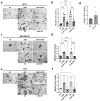
LDBK and BK, through B1 and B2 receptors, increase transwell migration in MCF-7, MDA-MB-231, and T47D cells. (a,c,e): the effects of LDBK and BK, and their antagonists, on transwell migration in MCF-7, MDA-MB-231, and T47D cells (pictures magnification corresponds to 400×); (b,d,f): the graphs show the fold change in stimulated migrating cells compared with the control. The number of migrating cells was normalized by the non-stimulated cells (NS), and (g) the graph shows the effect of the vehicle on the invasion of the MDA-MB-231 cells after 24 h of stimulus. Data are presented as the average ± standard error for at least three independent experiments. *** p < 0.001, and **** p < 0.0001. An ANOVA two-tailed test was used.
Figure 3
Figure 3
B1 and B2 receptors mediate the kinin-induced invasion of breast cancer cells. (a,c,e): the effects of LDBK and BK, and their antagonists, on Matrigel invasion in MCF-7, MDA-MB-231, and T47D cells (pictures magnification corresponds to 400×); (b,d,f): the graphs show the fold change in stimulated invasive cells compared with the control. The number of invasive cells was normalized by the non-stimulated cells (NS), and (g) the graph shows the effect of the vehicle on the invasion of MDA-MB-231 cells after 24 h of stimulus. Data are presented as the average ± standard error for at least three independent experiments. * p < 0.05, ** p < 0.01, *** p < 0.001, and **** p < 0.0001. An ANOVA two-tailed test was used.
Figure 4
Figure 4
Migration and invasion induced by kinin nonapeptides in breast cancer cells are mediated by Src and FAK. (a,c): transwell assay of T47D and MDA-MB-231 cells incubated with Src (PP2) and FAK (N14) inhibitors before the addition of kinins. Cells non-stimulated with agonists or inhibitors were considered controls (NS); (b,d): the graphs show the fold change in the stimulated T47D and MDA-MB-231 migrating cells compared with the non-stimulated cells. The number of migrating cells was normalized by the control cells; (e,g): Matrigel assay of T47D and MDA-MB-231 cells incubated with PP2 and N14 inhibitors plus LDBK or BK for 24 h. Cells without stimuli were considered controls (NS); and (f,h): the graphs show the fold change in the stimulated T47D and MDA-MB-231 invasive cells compared with the non-stimulated cells. The number of invasive cells obtained in each condition was normalized by the control cells. Pictures magnification corresponds to 400×. Data are presented as the average ± standard error for at least three independent experiments. * p < 0.05, ** p < 0.01, *** p < 0.001, and **** p < 0.0001. An ANOVA two-tailed test was used.
Figure 5
Figure 5
B1 and B2 antagonists impair tumor growth in vivo: (a) diagram of the protocol for breast cancer tumor formation based on the subcutaneous inoculation of MDA-MB-231 cells. Previously inoculated cells were treated for 24 h with B1 (AntB1) or B2 (AntB2) antagonists. Non-stimulated cells were used as controls; (b) tumor growth progression from the beginning to the 21st day; (c) tumor size at the end of the experiment (21st day); (d) representative pictures of tumor volume at different experimental conditions; (e) probability of the survival of mice under different conditions for 21 days; and (f) capacity of the proliferation of cells previously treated for 24 h with B1 and B2 antagonists. Data of six animals per group are presented as the average ± standard deviation. ** p < 0.01, *** p < 0.001, and **** p < 0.0001. A t-test was used.
Figure 6
Figure 6
The role of kinin receptors in breast cancer cell migration, invasion, and tumor growth in vivo. This figure illustrates the proposed mechanism by which B1 and B2 receptor activation leads to enhanced cell motility and invasiveness, while receptor antagonism hinders tumor progression: (a) activation of B1 and B2 receptors by LDBK and BK induces migration and invasion in breast cancer cells through the FAK–Src signaling pathway; and (b) in vivo antagonism of both B1 and B2 receptors impairs breast tumor growth, highlighting the therapeutic potential of receptor blockade in breast cancer treatment.

Similar articles

References

    1. Melzer C., Ohe J., Hass R. Breast Carcinoma: From Initial Tumor Cell Detachment to Settlement at Secondary Sites. BioMed Res. Int. 2017;2017:8534371. doi: 10.1155/2017/8534371. - DOI - PMC - PubMed
    1. Sung H., Ferlay J., Siegel R.L., Laversanne M., Soerjomataram I., Jemal A., Bray F. Global Cancer Statistics 2020: GLOBOCAN Estimates of Incidence and Mortality Worldwide for 36 Cancers in 185 Countries. CA Cancer J. Clin. 2021;71:209–249. doi: 10.3322/caac.21660. - DOI - PubMed
    1. Ingthorsson S., Briem E., Bergthorsson J.T., Gudjonsson T. Epithelial Plasticity During Human Breast Morphogenesis and Cancer Progression. J. Mammary Gland Biol. Neoplasia. 2016;21:139–148. doi: 10.1007/s10911-016-9366-3. - DOI - PMC - PubMed
    1. Fan T., Kuang G., Long R., Han Y., Wang J. The overall process of metastasis: From initiation to a new tumor. Biochim. Biophys. Acta (BBA) Rev. Cancer. 2022;1877:188750. doi: 10.1016/j.bbcan.2022.188750. - DOI - PubMed
    1. Seyfried T.N., Hyusentruyt L.C. On the Origin of Cancer Metastasis. Crit. Rev. Oncog. 2014;18:43–73. doi: 10.1615/CritRevOncog.v18.i1-2.40. - DOI - PMC - PubMed

MeSH terms

Grants and funding

LinkOut - more resources