Review
. 2024 Nov 1;25(21):11762.
doi: 10.3390/ijms252111762.

Electron Tomography as a Tool to Study SARS-CoV-2 Morphology

Affiliations
Review

Electron Tomography as a Tool to Study SARS-CoV-2 Morphology

Hong Wu et al. Int J Mol Sci. .

Abstract

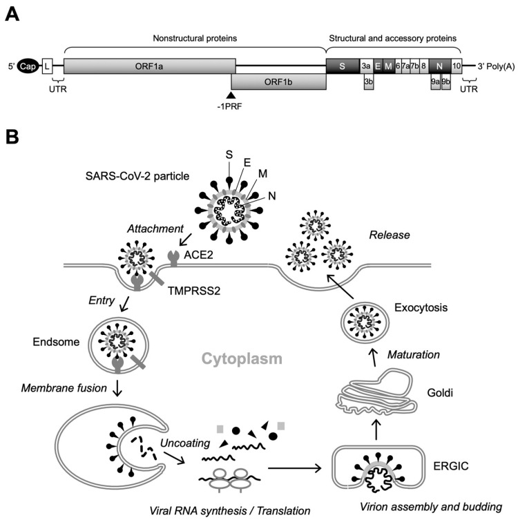
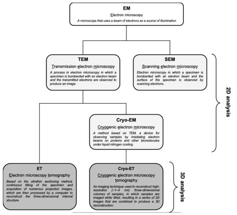
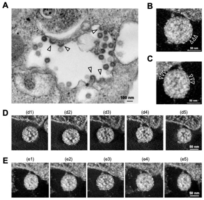
Severe acute respiratory syndrome coronavirus 2 (SARS-CoV-2), a novel betacoronavirus, is the causative agent of COVID-19, which has caused economic and social disruption worldwide. To date, many drugs and vaccines have been developed for the treatment and prevention of COVID-19 and have effectively controlled the global epidemic of SARS-CoV-2. However, SARS-CoV-2 is highly mutable, leading to the emergence of new variants that may counteract current therapeutic measures. Electron microscopy (EM) is a valuable technique for obtaining ultrastructural information about the intracellular process of virus replication. In particular, EM allows us to visualize the morphological and subcellular changes during virion formation, which would provide a promising avenue for the development of antiviral agents effective against new SARS-CoV-2 variants. In this review, we present our recent findings using transmission electron microscopy (TEM) combined with electron tomography (ET) to reveal the morphologically distinct types of SARS-CoV-2 particles, demonstrating that TEM and ET are valuable tools for visually understanding the maturation status of SARS-CoV-2 in infected cells. This review also discusses the application of EM analysis to the evaluation of genetically engineered RNA viruses.

Keywords: SARS-CoV-2; electron microscopy; electron tomography; morphogenesis; three-dimensional reconstruction; transmission electron microscopy; virus budding.


Conflict of interest statement

The authors declare no conflict of interest.

Figures

Figure 1
Figure 1
Genome structure and replication cycle of SARS-CoV-2. (A) Genomic organization. Sixteen NSPs are expressed as polyproteins from ORF1a and ORF1b and produced by internal cleavage by viral proteases, NSp3 (PLpro) and Nsp5 (Mpro), while four structural proteins (S, E, M, and N) are expressed from individual ORFs [9]. Interspersed ORFs in the 3’ portion of the genome encode accessory proteins (ORF3a, ORF3b, ORF6, ORF7a, ORF7b, ORF8, ORF9b, ORF9c, and ORF10), which have been reported to play key roles in viral pathogenesis [10]. There is a slippery sequence (programmed -1 ribosomal frameshifting [-1PRF]) site at the overlap of ORF1a and ORF3b, where a portion of the translating ribosomes slips back one nucleotide, resulting in the polyprotein synthesis of ORF1b [11]. Cap, 5’ cap structure; L, leader sequence; UTR, untranslated region. (B) Overview of SARS-CoV-2 replication. After binding of the S protein to cell-surface ACE2, the attached virion is internalized into the cell via endocytosis. Specific cleavage of the S protein by cellular proteases, including TMPRSS2, then triggers fusion between the viral and endosomal membranes. Following the uncoating of the incoming virion, polyproteins of ORF1a and ORF1b are translated from the released viral RNA, which in turn is cleaved to produce NSPs that form the RTC. The viral RNA amplification process occurs on the ER membrane and translated structural proteins translocate to the lumen of the ERGIC to assemble virus particles. Finally, infectious virions are released from infected cells by exocytosis.
Figure 2
Figure 2
The general principle of ET.
Figure 3
Figure 3
Types of EM techniques and their abbreviations. TEM provides high-resolution 2D images of ultrathin sections, which is useful for observing broader and more complex structures within a sample. ET is a TEM-based method that generates 3D reconstructions of a given area by tilting the sample and capturing multiple images. On the other hand, cryo-EM was developed for more detailed structural analysis, and cryo-ET is a specialized application of cryo-EM that reconstitutes cryo-EM data into a 3D structure. SEM is one of the electron microscopy techniques that is beyond the scope of this review.
Figure 4
Figure 4
Intracellular SARS-CoV-2 image obtained via TEM and ET. (A) Conventional TEM image of budding virus particles (arrowheads); (B,C) 3D ET images of budding virion in Y (B) and X (C) rotation. Arrowheads indicate S proteins on the surfaces of the virions. (D,E) Sequential 3D ET images in the Z-slice position obtained from two videos (d1–d5 and e1–e5). All panels are adapted with permission from [30]. Copyright 2022 Wu et al. https://creativecommons.org/licenses/by/4.0/ (accessed on 28 October 2024).
Figure 5
Figure 5
Distinct morphologies of SARS-CoV-2 observed in the vacuoles. (A) TEM image of hollow viral particles (white arrowheads in insert) budding within the intracellular vacuole observed near the nucleus. (B) Representative 3D ET images (in Y rotation) of SARS-CoV-2 particles inside the intracellular vacuole (left and middle panels) and on the surface membrane (right panel) of infected Calu-3 cells. (C,D) Z-slice position image (i.e., from c1 to c5 and d1 to d5) of hollow (C) and dense (D) particles budding within the vacuole. All panels are adapted with permission from [31]. Copyright 2024 Wu et al. https://creativecommons.org/licenses/by/4.0/ (accessed on 28 October 2024).

Similar articles

References

    1. Colantuoni A., Martini R., Caprari P., Ballestri M., Capecchi P.L., Gnasso A., Presti R.L., Marcoccia A., Rossi M., Caimi G. COVID-19 Sepsis and Microcirculation Dysfunction. Front. Physiol. 2020;11:747. doi: 10.3389/fphys.2020.00747. - DOI - PMC - PubMed
    1. Wu C., Chen X., Cai Y., Xia J., Zhou X., Xu S., Huang H., Zhang L., Zhou X., Du C., et al. Risk Factors Associated with Acute Respiratory Distress Syndrome and Death in Patients With Coronavirus Disease 2019 Pneumonia in Wuhan, China. JAMA Intern. Med. 2020;180:934–943. doi: 10.1001/jamainternmed.2020.0994. - DOI - PMC - PubMed
    1. WHO Coronavirus Disease (COVID-19) [(accessed on 1 August 2024)]. Available online: https://www.who.int/emergencies/diseases/novel-coronavirus-2019.
    1. WHO Director-General’s Opening Remarks at the Media Briefing—5 May 2023. [(accessed on 28 October 2024)]. Available online: https://www.who.int/director-general/speeches/detail/who-director-genera....
    1. Li X., Mi Z., Liu Z., Rong P. SARS-CoV-2: Pathogenesis, Therapeutics, Variants, and Vaccines. Front. Microbiol. 2024;15:1334152. doi: 10.3389/fmicb.2024.1334152. - DOI - PMC - PubMed

Supplementary concepts

LinkOut - more resources