. 2024 Nov 1;13(21):3080.
doi: 10.3390/plants13213080.

Induction and Characteristics of Callus Cultures of the Medicinal Plant Tussilago farfara L

Affiliations

Induction and Characteristics of Callus Cultures of the Medicinal Plant Tussilago farfara L

Monika Bojko et al. Plants (Basel). .

Abstract

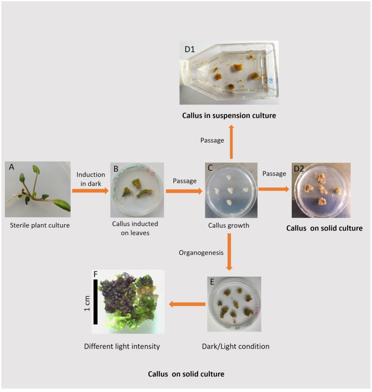
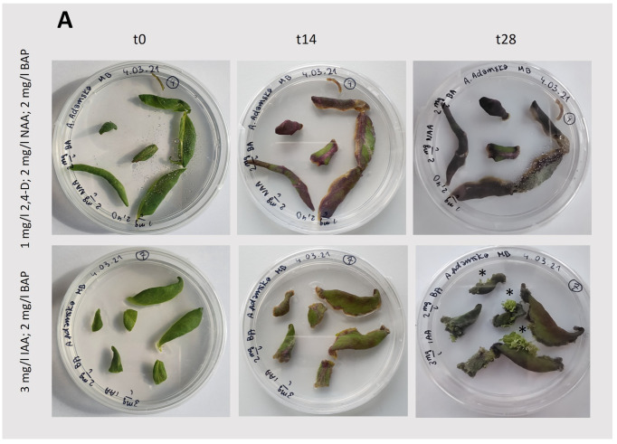
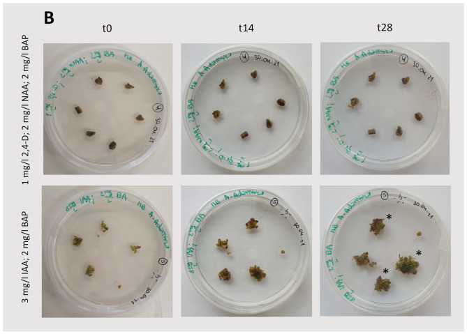
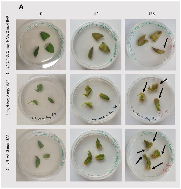
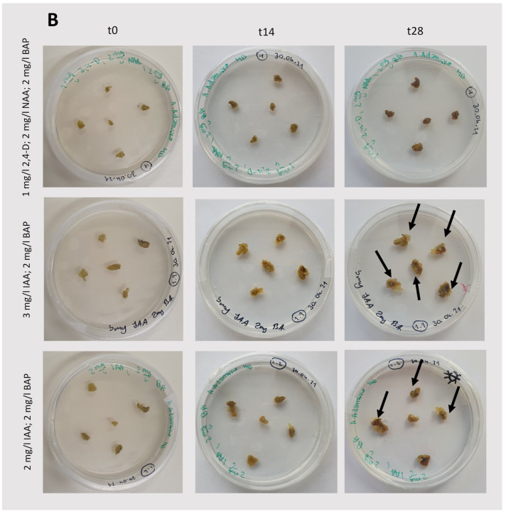
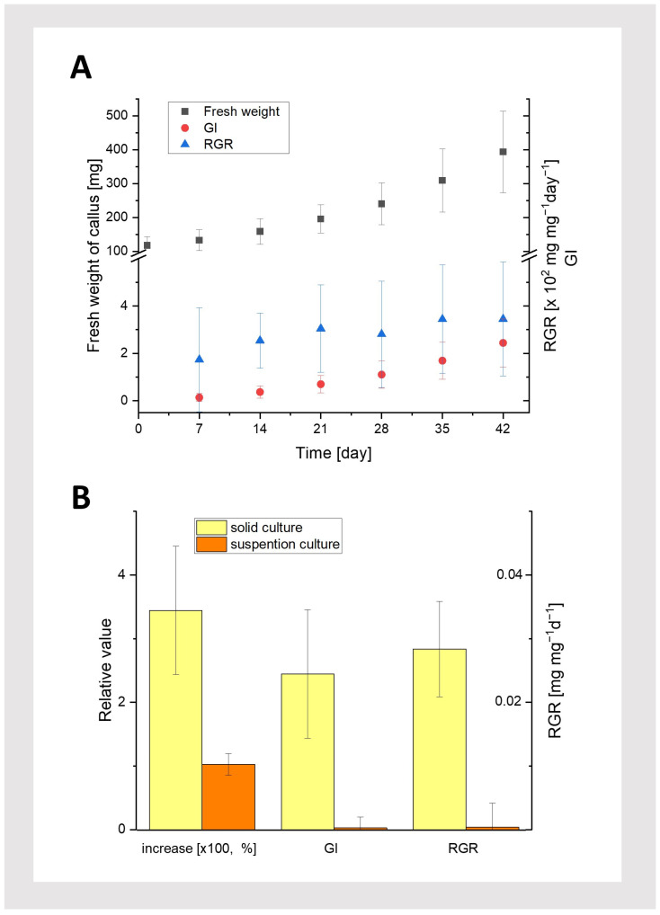
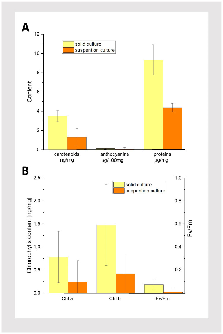
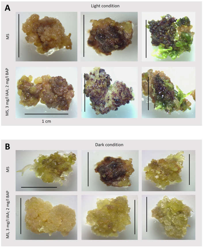
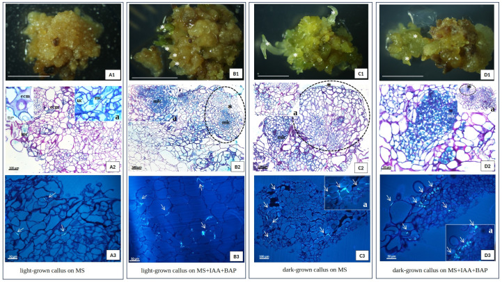
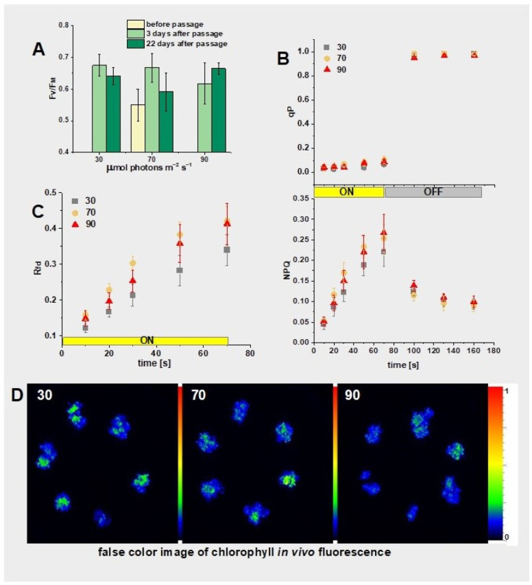
Tussilago farfara L. is a traditional medicinal plant valued for its potentially health-promoting metabolites. Its herbal raw material has been recognized and used since ancient times and continues to be widely used in traditional medicine. Introducing this plant species to in vitro cultivation is a challenging task, but once the protocol is developed, such cultures can provide an abundant and inexhaustible source of plant material. In this study, we report the successful induction and growth of vigorous T. farfara callus in vitro. Callus induction was achieved on MS solid media with the combination of indole-3-acetic acid (3 mg/L) and benzyl aminopurine (2 mg/L) in darkness, whereas it appeared inefficient under light conditions and in suspension culture. We present a detailed description of callus growth kinetics, morphological analysis, photosynthetic activity, and biochemical parameters (including protein content and photosynthetic pigments) supported by histological studies. Furthermore, we observed the potential for organogenesis and somatic embryogenesis. This method for the in vitro propagation of T. farfara, along with callus culture maintenance, offers a wide range of applications in pharmacy for the production of valuable metabolites. Moreover, it could benefit the environment by reducing the depletion of natural populations of this species and may serve as an alternative strategy for species conservation in light of global warming.

Keywords: carotenoids; chlorophyll; coltsfoot; fluorescence; histology; organogenesis.


Conflict of interest statement

The authors declare no conflicts of interest.

Figures

Figure 1
Figure 1
Tussilago farfara L. in its natural habitat (meadows near the Campus of the 600th Anniversary of the Jagiellonian University in Kraków (50°01′44,396″ N 19°54′17,496″ E; Poland). (A): Yellow inflorescences (capitula) visible in early spring, developing before the leaves, alongside thin, underground branched rhizomes. (B): Large, round, heart-shaped leaves with radial veins and crinkly, slightly toothed edges, documented in summer.
Figure 2
Figure 2
Schematic overview of the experiment, including illustrative examples of the results. Leaf explants were taken from a T. farfara plant, regenerated from a callus and grown in vitro (A), and used for the induction of a callus (B); A callus, separated from the leaf explants, is subcultured on solid medium (C); The callus is transferred to a suspension culture (D1) and on solid media (D2), with the addition of various hormones and grown in darkness. Finally, after 42 days of growth in darkness on solid media and in suspension culture, the callus tissue was taken for biochemical analyses (D1,D2). Additionally, stabilized callus cultures (C) were subcultured and grown under dark/light conditions and used for histochemical study (E); morphogenic callus from darkness (C), transferred to various light conditions (F).
Figure 3
Figure 3
Induction and growth of the callus under light conditions (75 μmol photons m−2 s −1; 14/10 (light/dark) photoperiod). (A): Callus induction from the leaf blade (Figure 2B). (B): Callus growth after initiation; t0—the day of callus passage, the starting point of observation; t14 and t28—observations after 14 and 28 days, respectively. In the case of the oldest cultures (t28), exemplary shoots are indicated by stars. The composition of the medium is specified in the figure.
Figure 3
Figure 3
Induction and growth of the callus under light conditions (75 μmol photons m−2 s −1; 14/10 (light/dark) photoperiod). (A): Callus induction from the leaf blade (Figure 2B). (B): Callus growth after initiation; t0—the day of callus passage, the starting point of observation; t14 and t28—observations after 14 and 28 days, respectively. In the case of the oldest cultures (t28), exemplary shoots are indicated by stars. The composition of the medium is specified in the figure.
Figure 4
Figure 4
Induction and growth of the callus under dark conditions. (A): Callus induction from the leaf blade (Figure 2B). (B): Callus growth after initiation; t0—the day of callus passage, the starting point of observation; t14 and t28—observations after 14 and 28 days, respectively. In the case of the oldest cultures (t28), exemplary callus is pointed by arrows. The composition of the medium is specified in the figure.
Figure 4
Figure 4
Induction and growth of the callus under dark conditions. (A): Callus induction from the leaf blade (Figure 2B). (B): Callus growth after initiation; t0—the day of callus passage, the starting point of observation; t14 and t28—observations after 14 and 28 days, respectively. In the case of the oldest cultures (t28), exemplary callus is pointed by arrows. The composition of the medium is specified in the figure.
Figure 5
Figure 5
Callus growth characteristics. (A): Growth kinetics (fresh weight), Growth Index (GI), and Relative Growth Rate (RGR; harvest interval: 7 days) of the callus grown under dark conditions on solid media (MS, 3 mg/L IAA; 2 mg/L BAP, 0.6% agar). (B): Increase in fresh weight, Growth Index (GI), and Relative Growth Rate (RGR) for the callus from solid and suspension cultures (MS, 3 mg/L IAA; 2 mg/L BAP) under dark conditions, measured on the 42nd day of culture. Average values and standard deviations are shown for n = 25 callus fragments.
Figure 6
Figure 6
Results of biochemical tests performed on dark-grown 42-day-old solid and suspension cultures of T. farfara. (A): Content of carotenoids, anthocyanins, and proteins. (B): Content of chlorophylls and FV/FM; n = 3, n represents individual pieces of callus.
Figure 7
Figure 7
Organogenesis, color, and structure of an exemplary callus grown for 42 days on different media. (A): callus grown under light conditions 75 μmol photons m−2 s −1; 14/10 (light/dark) photoperiod; (B): callus grown in dark conditions; Scale bar—1 cm.
Figure 8
Figure 8
Histological analysis of T. farfara callus sampling 42 days after subculture. (A): light-grown callus on MS, (B): light-grown callus on MS + 3 mg/L IAA + 2 mg/L BAP, (C): dark-grown callus on MS, (D): dark-grown callus on MS + 3 mg/L IAA + 2 mg/L BAP; (A2,B2,C2,D2)—Pass and Naphtol Blue Back staining, (A3,B3,C3,D3)—aniline blue staining; arrows—callose deposition, asterisk—regenerants, ecm—extra cellular matrix, mlc—meristematic like cells, uc—unfinished cytokinesis, mc—multinucleated cell; Scale bar of (A1,B1,C1,D1)—0.5 cm; light intensity—75 μmol photons m−2 s −1.
Figure 9
Figure 9
Photosynthetic parameters of the callus with adventitious shoots obtained from a chlorophyll in vivo fluorescence image. The culture was performed on MS, 3 mg/L IAA; 2 mg/L BAP under different light intensities (30, 70, and 90 μmol photons m−2 s−1; labeled as 30, 70, and 90, respectively). (A): FV/FM; (B): Coefficient of photochemical quenching (qP) and nonphotochemical quenching (NPQ) measured for 22 days after passage; (C): Vitality index (Rfd) measured in parallel to qP and NPQ; (AC) Average values and standard deviations are shown for n = 6; (B,C) The yellow and gray rectangles indicate turning on and off the actinic light (600 μmol photons m−2 s−1), respectively. (D): False color fluorescence image (FM) of a 28-day-old callus grown under different light intensities; see Section 3 for further explanations.
Figure 10
Figure 10
Sample close-up images of callus cultured for 22 days on MS, 3 mg/L IAA; 2 mg/L BAP under light with different intensities, as shown in the figure (left side), along with the corresponding false color chlorophyll fluorescence image, corresponding to FM and induced with a saturating pulse of white light (right side); see Section 3 for details.

Similar articles

Cited by

References

    1. [(accessed on 19 September 2024)]. Available online: https://powo.science.kew.org/taxon/urn:lsid:ipni.org:names:256904-1.
    1. Tobyn G., Denham A., Whitelegg M., editors. Medical Herbs. Churchill Livingstone; Edinburgh, UK: 2011. CHAPTER 30—Tussilago farfara, coltsfoot; pp. 317–326. - DOI
    1. Matuszkiewicz W. Przewodnik do Oznaczania Zbiorowisk Roślinnych Polski. Wydawnictwo Naukowe PWN; Warszawie, Poland: 2006.
    1. Chen S., Dong L., Quan H., Zhou X., Ma J., Xia W., Zhou H., Fu X. A review of the ethnobotanical value, phytochemistry, pharmacology, toxicity and quality control of Tussilago farfara L. (coltsfoot) J. Ethnopharmacol. 2021;267:113478. doi: 10.1016/j.jep.2020.113478. - DOI - PMC - PubMed
    1. Adamczak A., Opala B., Gryszczyńska A., Buchwald W. Content of pyrrolizidine alkaloids in the leaves of coltsfoot (Tussilago farfara L.) in Poland. Acta Soc. Bot. Pol. 2013;82:289–293. doi: 10.5586/asbp.2013.028. - DOI

LinkOut - more resources