Review
. 2025 Feb;80(2):408-422.
doi: 10.1111/all.16382. Epub 2024 Nov 9.

Biologics and airway remodeling in asthma: early, late, and potential preventive effects

Affiliations
Review

Biologics and airway remodeling in asthma: early, late, and potential preventive effects

G Varricchi et al. Allergy. 2025 Feb.

Abstract

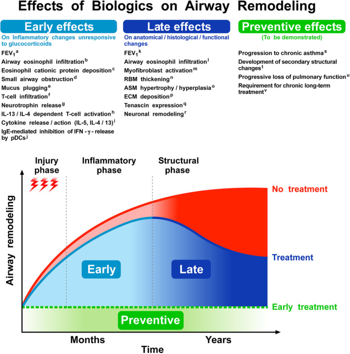
Although airway remodeling in severe and/or fatal asthma is still considered irreversible, its individual components as a cause of clinical symptoms and/or lung function changes remain largely unknown. While inhaled glucocorticoids have not consistently been shown to affect airway remodeling, biologics targeting specific pathways of airway inflammation have been shown to improve lung function, mucus plugging, and airway structural changes that can exceed those seen with glucocorticoids. This superiority of biologic treatment, which cannot be solely explained by insufficient doses or limited durations of glucocorticoid therapies, needs to be further explored. For this field of research, we propose a novel classification of the potential effects of biologics on airway remodeling into three temporal effects: early effects (days to weeks, primarily modulating inflammatory processes), late effects (months to years, predominantly affecting structural changes), and potential preventive effects (outcomes of early treatment with biologics). For the identification of potential preventive effects of biologics, we call for studies exploring the impact of early biological treatment on airway remodeling in patients with moderate-to-severe asthma, which should be accompanied by a long-term evaluation of clinical parameters, biomarkers, treatment burden, and socioeconomic implications.

Keywords: asthma treatment; biologics; remodeling.


Conflict of interest statement

GV reports research support from AstraZeneca. RP has no potential conflicts of interest to declare. ML has received consulting fees or honoraria, or both for lectures from ALK, Allergopharma, AstraZeneca, Berlin‐Chemie, Boehringer Ingelheim, Chiesi, GSK, HAL Allergy, Leti, Novartis, MSD, Sanofi, and TEVA; and grants for research or clinical trials, or both from Deutsche Forschungsgemeinschaft, AstraZeneca, and GSK. GB reports personal fees from Astra Zeneca, personal fees from Boehringer‐Ingelheim, personal fees from Chiesi, personal fees from GSK, personal fees from Novartis, personal fees from Sanofi, grants from MSD, outside the submitted work. G.W.C. has received consulting fees or honoraria, or both for lectures from AstraZeneca, Chiesi, Novartis, Sanofi, Menarini, Stallergenes Greer, GSK, and HAL Allergy. FB has been on the scientific Board for AZ, BI, Chiesi, GSK, Menarini group, Sanofi and P&G; received honoraria from AZ, BI, Chiesi, GSK, Menarini group and Sanofi. JCV is a full time employee of the University of Rostock as a full time professor and chair of the Departments of Pneumology and Intensive Care Medicine has given independent advice, lectured for and received honoraria from AstraZeneca, Avontec, Bayer, Bencard, Bionorica, Boehringer‐Ingelheim, Chiesi, Essex/Schering‐Plough, GSK, Janssen‐Cilag, Leti, MEDA, Merck, MSD, Mundipharma, Novartis, Nycomed/Altana, Pfizer, Revotar, Sandoz‐Hexal, Stallergens, TEVA, UCB/Schwarz‐Pharma, Zydus/Cadila, has participated in advisory boards for Avontec, Boehringer‐Ingelheim, Chiesi, Essex/Schering‐Plough, GSK, Janssen‐Cilag, MEDA, MSD, Mundipharma, Novartis, Regeneron, Revotar, Roche, Sanofi‐Aventis, Sandoz‐Hexal, TEVA, UCB/Schwarz‐Pharma and has received research grants from the Deutsche Forschungsgesellschaft, Land Mecklenburg‐Vorpommern, GSK, MSD. G.W.C. received honoraria for lectures, presentations, speakers from AstraZeneca, GSK, Novartis, Sanofi, Stallergenes, Greer, Hal Allergy, Menarini, Chiesi, Mylan, Valeas, Faes.

Figures

FIGURE 1
FIGURE 1
(A) FEV1 (1.3 L [49% predicted]) in a patient with severe asthma and repeated severe OCS‐dependent exacerbations despite maintenance treatment with oral prednisolone (5 mg/day per os) prior to biologics. (B) The red lines show the FEV1 improvement (1.6 L [61% predicted]) after seven courses of mepolizumab (100 mg s.c./4 weeks) without prednisolone. (C) The red lines show FEV1 improvement [FEV1 2.8 L (105% predicted)] after five courses of reslizumab (3 mg/kg IV/4 weeks) without prednisolone. F/V ex, flow/volume during expiration; F/V in, flow/volume during inspiration; ITGV, intrathoracic gas volume; RV, residual volume; TLC, total lung capacity; Vol, volume.
FIGURE 2
FIGURE 2
The figure schematically illustrates the complex mechanisms involved in airway remodeling (AiRem) in asthma. The figure also highlights the effects of various biological therapies in modulating these processes. The left side of the figure shows a normal airway compared to an asthmatic airway, which is characterized by inflammatory and structural changes such as epithelial damage, goblet cell metaplasia, mucus plugging, reticular basement thickening (RBM), airway smooth muscle (ASM) proliferation, subepithelial and peribronchial fibrosis, and increased angiogenesis. Several environmental factors (e.g., allergens, smoke, pollutants, microbial compounds, and nanoparticles) cause epithelial damage, leading to the release of alarmins (i.e., TSLP, IL‐33, and IL‐25). These cytokines activate a broad spectrum of innate (e.g., ILC2s) and adaptive (e.g., Th2 cells) immune cells, which promote airway inflammation and remodeling through the production of IL‐4, IL‐5, and IL‐13. TSLP plays a central role in AiRem by activating dendritic cells, eosinophils, mast cells, and human lung macrophages., TSLP also stimulates lung fibroblasts to produce extracellular matrix (ECM) components, promoting peribronchial fibrosis. In addition, TSLP induces the release of vascular endothelial growth factor (VEGF)‐A, the most potent angiogenic factor, from lung macrophages, and triggers epithelial‐mesenchymal transition (EMT). Tezepelumab, a human monoclonal antibody (mAb) that blocks TSLP, interferes with the activation of upstream and downstream inflammatory pathways, which contribute to goblet cell hyperplasia, mucus plugging, EMT, and angiogenesis. Dupilumab, an IL‐4Rα mAb antagonist, blocks both IL‐4 and IL‐13 signaling. IL‐4 promotes ASM proliferation by increasing Actin and collagen synthesis and enhancing TGF‐β release from the airway epithelium., IL‐13 disrupts epithelial integrity and promotes TGF‐β release from airway epithelial cells, stimulating airway fibrosis through the action of matrix metalloproteinases (MMPs). IL‐13 also enhances goblet cell metaplasia, contributing to mucus production and airway obstruction., , Dupilumab reduces mucus hypersecretion and goblet cell metaplasia and modulates ASM responses. Mepolizumab and reslizumab block IL‐5, which plays a critical role in subepithelial and peribronchial fibrosis by recruiting and activating eosinophils, which are major sources of TGF‐β., Mepolizumab reduces ASM proliferation and prevents IL‐5‐mediated epithelial damage and fibrosis., Benralizumab, by targeting IL‐5Rα, induces eosinophil depletion, thereby mitigating eosinophil‐driven AiRem. Benralizumab also reduces mucus plugging and ASM proliferation. Omalizumab, which targets IgE, reduces RBM thickening and fibronectin deposits in the bronchial mucosa. Itepekimab and Tozorakimab, interfering with IL‐33 signaling, may reduce AiRem by inhibiting TGF‐β pathways, although their role in clinical practice is still being evaluated. IL‐33 increases the production of collagen and fibronectin‐1 in lung fibroblasts, contributing to subepithelial and peribronchial fibrosis. Red lines indicate inflammatory mechanisms targeted by biologics in early intervention, while blue lines represent the modulation of structural changes in the late phase of treatment. ASM, airway smooth muscle; Col‐1, collagen‐1; EMT, epithelial‐mesenchymal transition; IL, interleukin; IL‐5R, interleukin‐5 receptor; ILC2, innate lymphoid cell type 2; LTC4, leukotriene C4; MMP, matrix metalloproteinase; RBM, reticular basement thickness; ROS, reactive oxygen species; TGF, transforming growth factor; TH, T helper lymphocyte; TSLP, thymic stromal lymphopoietin; TSLPR, thymic stromal lymphopoietin receptor.
FIGURE 3
FIGURE 3
Early effects: Evidence of improvements in lung function and reduction in inflammation shortly after biologics' initiation illustrate the rapid benefits of targeting specific inflammatory pathways (a, , , , , , , , ; b; c; d; e; f; g; h, ; i; j 135 ). Late effects: Analogous to observed outcomes in conditions like atopic dermatitis, and eosinophilic oesophagitis, biologics may also impact long‐term structural changes within the airways, suggesting a potential for reversing or ameliorating established AiRem (k, , , , , , ; l; m; n, ; o, , ; p; q; r 140 ). Potential preventive effects: This represents an emerging research domain, emphasizing the potential of biologics to prevent the onset or progression of AiRem. Although empirical evidence is presently limited in this area, it is identified as a vital avenue for future research aimed at elucidating the comprehensive effects of biologics on the asthma disease trajectory (s; t; u; v 143 ). It is important to note that the studies regarding the early and late effects of biologics are essentially based on adults with severe asthma. Further research is needed to evaluate early, late, and potential preventive effects of biologics in pediatric cohorts. ASM, airway smooth muscle; ECM, collagen and extracellular matrix; FEV1, forced expiratory volume in 1 s; IFN, interferon; IL, interleukin; pDC, plasmacytoid dendritic cell; RBM, reticular basement membrane.

Similar articles

Cited by

References

    1. Lommatzsch M, Brusselle GG, Canonica GW, et al. Disease‐modifying anti‐asthmatic drugs. Lancet. 2022;399:1664‐1668. doi:10.1016/S0140-6736(22)00331-2 - DOI - PubMed
    1. Bousquet J, Chanez P, Lacoste JY, et al. Asthma: a disease remodeling the airways. Allergy. 1992;47:3‐11. doi:10.1111/j.1398-9995.1992.tb02242.x - DOI - PubMed
    1. Vignola AM, Chanez P, Bonsignore G, Godard P, Bousquet J. Structural consequences of airway inflammation in asthma. J Allergy Clin Immunol. 2000;105:S514‐S517. doi:10.1016/s0091-6749(00)90053-8 - DOI - PubMed
    1. Green FH, Williams DJ, James A, et al. Increased myoepithelial cells of bronchial submucosal glands in fatal asthma. Thorax. 2010;65:32‐38. doi:10.1136/thx.2008.111435 - DOI - PubMed
    1. James AL, Elliot JG, Jones RL, et al. Airway smooth muscle hypertrophy and hyperplasia in asthma. Am J Respir Crit Care Med. 2012;185:1058‐1064. doi:10.1164/rccm.201110-1849OC - DOI - PubMed

MeSH terms