. 2025 Mar;14(6):e2403119.
doi: 10.1002/adhm.202403119. Epub 2024 Nov 9.

Minimally Invasive Syringe-Injectable Hydrogel with Angiogenic Factors for Ischemic Stroke Treatment

Affiliations

Minimally Invasive Syringe-Injectable Hydrogel with Angiogenic Factors for Ischemic Stroke Treatment

Donggue Kim et al. Adv Healthc Mater. 2025 Mar.

Abstract

Ischemic stroke (IS) accounts for most stroke incidents and causes intractable damage to brain tissue. This condition manifests as diverse aftereffects, such as motor impairment, emotional disturbances, and dementia. However, a fundamental approach to curing IS remains unclear. This study proposes a novel approach for treating IS by employing minimally invasive and injectable jammed gelatin-norbornene nanofibrous hydrogels (GNF) infused with growth factors (GFs). The developed GNF/GF hydrogels are administered to the motor cortex of a rat IS model to evaluate their therapeutic effects on IS-induced motor dysfunction. GNFs mimic a natural fibrous extracellular matrix architecture and can be precisely injected into a targeted brain area. The syringe-injectable jammed nanofibrous hydrogel system increased angiogenesis, inflammation, and sensorimotor function in the IS-affected brain. For clinical applications, the biocompatible GNF hydrogel has the potential to efficiently load disease-specific drugs, enabling targeted therapy for treating a wide range of neurological diseases.

Keywords: angiogenesis; electrospinning; gelatin‐norbornene; ischemic stroke; sensorimotor function.


Conflict of interest statement

The authors declare no conflict of interest.

Figures

Figure 1
Figure 1
Schematic illustration of the experiments. In an animal IS model, the injection of a GNF/GF hydrogel (with a structure similar to the fibrous ECM) into the motor cortex enhanced angiogenesis and sensorimotor function.
Figure 2
Figure 2
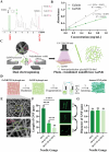
Overall process to fabricate injectable nanofibrous GelNB hydrogels. Degree of substitution of GelNB measured by A) 1H NMR and B) TNBSA assay. The obtained NMR peaks were analyzed by comparing the norbornene double bond signals (left, green) with the methyl proton signals (right, pink). The TNBSA assay was performed by comparing the slopes of the equations for Gelatin and GelNB. C) Illustration of dual electrospinning and photocrosslinking processes. The GelNB and PEO precursors were electrospun at a ratio of 4:6 and then exposed to UV light to photocrosslink the GelNB nanofibers (PEO nanofibers in the GelNB/PEO mat were not crosslinked). D) Illustration of the fabrication process after dual electrospinning. The photocrosslinked fibrous GelNB/PEO hydrogel mats were hydrated in PBS to remove the sacrificial PEO nanofibers. Then, pure GelNB hydrogel mats were fragmented by repeated needle pumping from 18 G to 26 G. The GNF solution was centrifuged to obtain jammed GNF pellet. E) Representative SEM images of the dual electrospun GelNB/PEO. The thicker fibers are GelNB and the thinner fibers are PEO. The yellow box represents the region where the magnified image below was obtained (×10). Scale bar: 10 µm (above), 3 µm (below). F) Length of GNFs at each step of needle pumping. Needles with a smaller inner diameter gave shorter GNFs. Scale bar: 10 µm (n = 30, p < 0.005). G) Diameter of the GNFs at each step of needle pumping. The fragmentation steps did not affect the diameter of the GNFs, which were all ≈800 nm (n = 50, p = 0.77). The numbers above each bar give the sample size (n) for each group. p values were determined by one‐way analysis ANOVA with Bonferroni's post‐hoc test in the three groups (needle gauge). All data are presented as the mean ± the standard error of the mean (SEM). *p< 0.1, **p< 0.05, ***p< 0.01, ****p< 0.005.
Figure 3
Figure 3
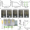
Rheological characteristics and precise volume control of the GNF hydrogel. Rheological characteristics of GNFs showing A) decreasing viscosity with increasing shear rate (0–50 1/s), B) shear‐yielding with increasing strain (0.037–1000 %, 1 Hz), and C) shear‐thinning and self‐healing during low‐strain (1 %, unshaded regions) and high‐strain (500 %, shaded regions) cycles at 1 Hz. D) Images captured during injection, needle translation to the left, and breakage of the GNF hydrogel with/without pressure. The yellow arrows indicate the height and width of the injected strand under pressure, and the yellow circles indicate the breaking and stacking of the strands when the pressure on the piston is removed. Each box in the grid has edges of 1 cm. E) Photographs showing the injectability of the GNF hydrogel as the sub‐microscale GNFs are smaller than the ID of the 30 G needle (0.16 mm). In contrast, bulk hydrogel and granular hydrogels samples cannot be injected because of their larger components. The red circles indicate the joints between the syringe and needle, through which the hydrogels need to pass during injection. The yellow box indicates the moment of injection and stacking of the GNF hydrogel. F) Schematic of the method used to precisely inject 1 µL doses of the GNF hydrogel (n = 7, p < 0.005) using a syringe pump at 1 µL min−1 with a 0.13 mm spacer. The numbers above each dot represent the sample size (n). All data are presented as the mean ± SEM.
Figure 4
Figure 4
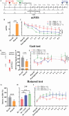
Sensorimotor function alleviation after injection of GNF/GF hydrogel. A) Overall timeline of behavior evaluations. To examine the neurotherapeutic effect of the injectable, jammed GNF/GF hydrogel on IS, we injected PBS, GNF, or GNF/GF into the motor cortex of an IS animal model, which was followed by behavior tests such as mNSS, gait test, and rotarod test for 8 weeks. mNSS tests implemented for the IS models B) pre‐IS and post‐IS (n = 20, p < 0.005) and C) post‐IS and post‐injection, measured every week for 8 weeks. The IS + GNF (n = 7) and IS + GNF/GF (n = 7) groups showed a reduced mNSS compared to the IS + PBS group (n = 6). D) Schematic describing the swing speeds of intact and lesioned hind paws, which were determined by the duration of each step (time between the paw leaving the ground and then landing). Measured LH swing speeds of the IS models showed E) a significant decrease in the Post‐IS group compared to the Pre‐IS group (n = 12, p < 0.005) and F) a significantly faster speed for the IS + GNF/GF group (n = 6) compared to the IS + PBS (n = 6) and IS + GNF (n = 7) groups over the 8 weeks post‐injection. The latency to falls determined by the rotarod test of the IS models showed a G) significantly lower value for the Post‐IS group compared to the Pre‐IS group (n = 20, p < 0.005). H) IS + GNF (n = 7) and IS + GNF/GF groups (n = 6) showed significantly higher latency compared to the IS + PBS group (n = 6) at week 3 (p = 0.005). I) Latency to falls measured at Post‐IS and Post‐injection states and weekly for 8 weeks after MCAO induction (IS + PBS: n = 6; IS + GNF: n = 7; IS + GNF/GF: n = 6). The number above each bar represents the sample size (n) for each group. The p values of the behaviors over time were determined by two‐way repeated measures ANOVA with a Bonferroni's post‐hoc test, one‐way ANOVA with a Bonferroni's post‐hoc test for the three groups (weekly tests), and a two‐tailed paired t‐test between Pre‐IS and Post‐IS groups. All data are presented as the mean ± SEM. *p< 0.1, **p< 0.05, ***p< 0.01, ****p< 0.005.
Figure 5
Figure 5
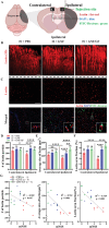
Angiogenesis induced by the GNF/GF hydrogel. A) Schematic of the brains of the IS models showing the injection point (green dots, AP ± 1 mm, ML ‐1 mm, DV +1 mm), analysis area (black boxes, C: contralateral, I: ipsilateral), staining dyes (Lectin: blood vessels; DAPI: nucleus; FITC‐Dextran: GNFs) and infarction region (gray shaded area). B) Lectin (far‐red) labeling of perfusable blood vessels was visualized using fluorescence 3D images of the IS models 8 weeks after injection with PBS, GNF, or GNF/GF. Scale bar: 200 µm. C) DAPI (blue), lectin (far‐red), and GNFs (FITC‐dextran: green) were visualized with fluorescence 2D images of sliced brains obtained 8 weeks after the injection. Representative images of lectin alone (top row) and the merged signals of lectin, DAPI, and FITC‐dextran (bottom row) in the ipsilateral region 8 weeks after injection of PBS, GNF, and GNF/GF groups. Scale bar: 200 µm. D) Number of lectic particles, E) average size of lectin areas, and F) lectin area fraction measured in the contralateral and ipsilateral hemisphere of the IS + PBS (n = 20), IS + GNF (n = 22), and IS + GNF/GF (n = 18) groups. G) Scatter plots showing the inverse correlations between mNSS and the number of lectin particle (left), average size of lectin areas (middle), and lectin area fraction (right). Each data point represents an individual animal. The number above each bar represents the sample size (n) for each group. The p values were determined by one‐way ANOVA with a Bonferroni's post‐hoc test for the three ipsilateral groups, and two‐tailed independent samples t‐test were used to compare between contralateral and ipsilateral hemisphere data from the same group. All data are presented as the mean ± SEM. *p< 0.1, **p< 0.05, ***p< 0.01, ****p< 0.005.

Similar articles

Cited by

References

    1. Tsao C. W., Aday A. W., Almarzooq Z. I., Anderson C. A. M., Arora P., Avery C. L., Baker‐Smith C. M., Beaton A. Z., Boehme A. K., Buxton A. E., Commodore‐Mensah Y., Elkind M. S. V., Evenson K. R., Eze‐Nliam C., Fugar S., Generoso G., Heard D. G., Hiremath S., Ho J. E., Kalani R., Kazi D. S., Ko D., Levine D. A., Liu J., Ma J., Magnani J. W., Michos E. D., Mussolino M. E., Navaneethan S. D., Parikh N. I., et al., Circulation 2023, 147, e93. - PubMed
    1. Donkor E. S., Stroke Res. Treat 2018, 2018, 3238165. - PMC - PubMed
    1. Kumar S., Selim M. H., Caplan L. R., Lancet Neurol. 2010, 9, 105. - PubMed
    1. Catanese L., Tarsia J., Fisher M., Circ. Res. 2017, 120, 541. - PubMed
    1. Zhang Z. G., Zhang L., Jiang Q., Zhang R., Davies K., Powers C., Bruggen N. V., Chopp M., J. Clin. Invest. 2000, 106, 829. - PMC - PubMed