

Red light-emitting diode on skin healing: an in vitro and in vivo experimental study
- PMID: 39521711
- PMCID: PMC11745292
- DOI: 10.1016/j.abd.2024.02.008
Red light-emitting diode on skin healing: an in vitro and in vivo experimental study
Abstract
Background: The clinical advantages of light-emitting diode (LED) therapy in skin healing and its underlying mechanism remain subjects of ongoing debate.
Objective: This study aims to explore the impact of LED therapy on normal skin keratinocytes (HaCaT) and in the repair of full-thickness dorsal wounds in Wistar rats.
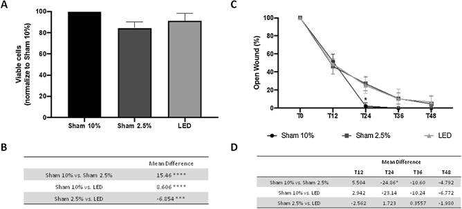
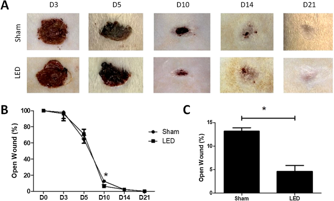
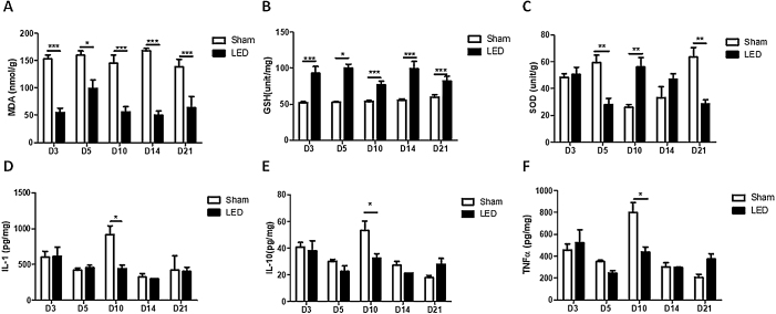
Methods: HaCaT cell viability (SRB assay) and migration (scratch assay) were assessed under LED therapy, comparing stress conditions (2.5% FBS) with sham irradiation and optimal conditions (10% FBS). In vivo, 50 rats with induced wounds were divided into Sham and LED (daily treatment) groups. Euthanasia occurred at 3, 5, 10, 14, and 21 days for clinical, morphological, oxidative stress (MDA, SOD, and GSH), and cytokine analyses (IL-1β, IL-10, TNF-α).
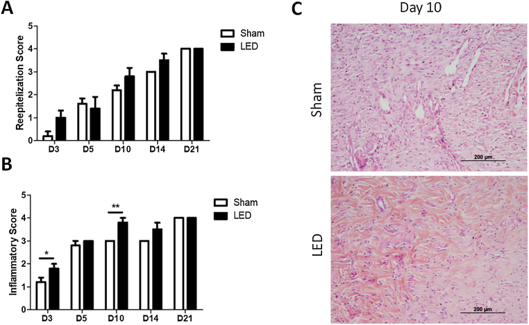
Results: LED therapy significantly enhanced keratinocytes viability compared to sham irradiation, with minimal impact on cell migration. Clinical benefits were prominent on day 10, influencing inflammation progression and resolution on days 3 and 10. Re-epithelization remained unaffected. Reduced MDA and increased GSH levels were observed throughout, while SOD levels varied temporally. Notably, on day 10, LED significantly decreased IL-1β, IL-10, and TNF-α.
Study limitations: Although translational, clinical trial confirmation of observed benefits is warranted.
Conclusions: LED therapy expedites cutaneous healing in the experimental model, primarily modulating inflammation and enhancing antioxidant activity.
Keywords: Cell culture techniques; Cytokines; Dermatology; Low-level light therapy; Models, animal; Oxidative stress.
Copyright © 2024 Sociedade Brasileira de Dermatologia. Published by Elsevier España, S.L.U. All rights reserved.
Conflict of interest statement
Conflicts of interest None declared.
Figures
Similar articles
-
Defocused high-power diode laser accelerates skin repair in a murine model through REDOX state modulation and reepithelization and collagen deposition stimulation.J Photochem Photobiol B. 2021 Dec;225:112332. doi: 10.1016/j.jphotobiol.2021.112332. Epub 2021 Oct 9. J Photochem Photobiol B. 2021. PMID: 34653929
-
Green light emitting diodes accelerate wound healing: characterization of the effect and its molecular basis in vitro and in vivo.Wound Repair Regen. 2012 Mar-Apr;20(2):226-35. doi: 10.1111/j.1524-475X.2012.00771.x. Wound Repair Regen. 2012. PMID: 22380691
-
Does blue light restore human epidermal barrier function via activation of Opsin during cutaneous wound healing?Lasers Surg Med. 2019 Apr;51(4):370-382. doi: 10.1002/lsm.23015. Epub 2018 Aug 31. Lasers Surg Med. 2019. PMID: 30168605
-
Effect of NASA light-emitting diode irradiation on wound healing.J Clin Laser Med Surg. 2001 Dec;19(6):305-14. doi: 10.1089/104454701753342758. J Clin Laser Med Surg. 2001. PMID: 11776448 Review.
-
The role of light emitting diode in wound healing: A systematic review of experimental studies.Cell Biochem Funct. 2024 Jul;42(5):e4086. doi: 10.1002/cbf.4086. Cell Biochem Funct. 2024. PMID: 38956862
References
-
- Nussbaum S.R., Carter M.J., Fife C.E., DaVanzo J., Haught R., Nusgart M., et al. An economic evaluation of the impact, cost, and medicare policy implications of chronic nonhealing wounds. Value Health. 2018;21:27–32. - PubMed
-
- Mármora B.C., Brochado F.T., Schmidt T.R., Santos L.G., Araújo A.A., Medeiros C.A.C.X., et al. Defocused high-power diode laser accelerates skin repair in a murine model through REDOX state modulation and reepithelization and collagen deposition stimulation. J Photochem Photobiol B. 2021;225 - PubMed
MeSH terms
Substances
LinkOut - more resources
Full Text Sources
