

Case Reports
doi: 10.1016/j.abd.2024.03.012.
Epub 2024 Nov 8.
Dermatofibroma: clinicopathological analysis of 239 cases
Affiliations
- PMID: 39521712
- PMCID: PMC11745288
- DOI: 10.1016/j.abd.2024.03.012
Item in Clipboard
Case Reports
Dermatofibroma: clinicopathological analysis of 239 cases
An Bras Dermatol.
2025 Jan-Feb.
No abstract available
Conflict of interest statement
Conflicts of interest None declared.
Figures
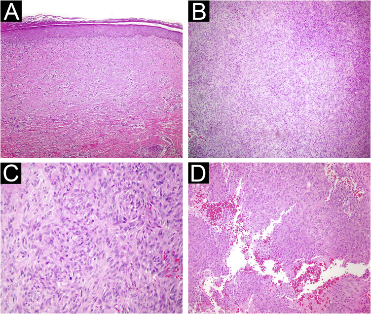
(A) Atrophic dermatofibroma. The lesion is hypocellular, and exhibits marked hyalinization. (Hematoxylin & eosin, ×200) (B) Cellular dermatofibroma with noticeable cellularity. (Hematoxylin & eosin, ×100) (C) High magnification of cellular dermatofibroma showing prominent mitotic figures. (Hematoxylin & eosin, ×400) (D) Aneurysmal fibrous histiocytoma. Cystic spaces filled with blood are observed within the lesion. (Hematoxylin & eosin, ×200).
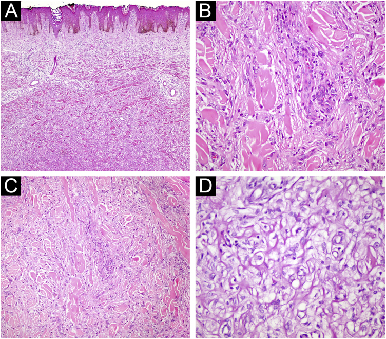
(A) Fibrous histiocytoma with atypical features. Marked nuclear pleomorphism is evident (Hematoxylin & eosin, ×200). (B) Fibrous histiocytoma with atypical features. Bizarre nucleated cells are observed (Hematoxylin & eosin, ×400). (C) Lipidized fibrous histiocytoma. At low magnification, significant hyalinization is noticeable in the lesion (Hematoxylin & eosin, ×40) (D) Foamy cytoplasmic appearance in the cytoplasm of lesional cells (Hematoxylin & eosin, ×400).
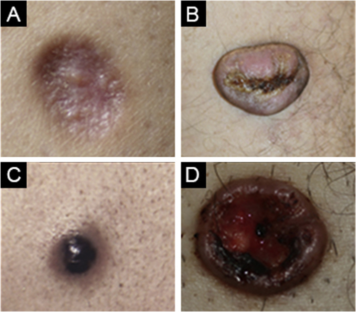
(A) Atrophic dermatofibroma. (B) Cellular dermatofibroma. (C) Aneurysmal dermatofibroma. (D) Lipidized dermatofibroma.
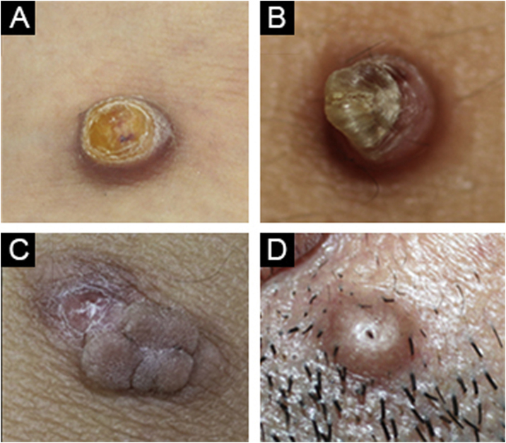
(A) Cellular dermatofibroma case with pre-diagnosis of eccrine poroma. (B) Common dermatofibroma case with a pre-diagnosis of keratoacanthoma. (C) Cellular dermatofibroma case with a pre-diagnosis of irritated skin-tag. (D) Common dermatofibroma case with a pre-diagnosis of trichofolliculoma.
References
-
- Felty C.C., Linos K. Epithelioid fibrous histiocytoma: a concise review. Am J Dermatopathol. 2019;41:879–883. - PubMed
-
- Lee W.J., Jung J.M., Won C.H., Chang S.E., Choi J.H., Moon K.C., et al. Clinical and histological patterns of dermatofibroma without gross skin surface change: a comparative study with conventional dermatofibroma. Indian J Dermatol Venereol Leprol. 2015;81:263–269. - PubMed
-
- Nabatanzi A., Male M., Qu X.Y., Li Y.Q., Meng X., Di W.S., et al. Aneurysmal fibrous histiocytoma: clinicopathology analysis of 30 cases of a rare variant of cutaneous fibrohistiocytoma. Curr Med Sci. 2019;39:134–137. - PubMed
Publication types
LinkOut - more resources
Full Text Sources
