. 2025 Jan 1;40(1):119-137.
doi: 10.1093/humrep/deae247.

Upregulated let-7 expression in the follicular fluid of patients with endometriomas leads to dysfunction of granulosa cells through targeting of IGF1R

Affiliations

Upregulated let-7 expression in the follicular fluid of patients with endometriomas leads to dysfunction of granulosa cells through targeting of IGF1R

Libing Shi et al. Hum Reprod. .

Abstract

Study question: What molecular mechanisms underlie the decline in ovarian reserve as the number and quality of oocytes decrease in patients with ovarian endometriomas (OEM)?

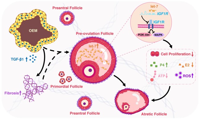
Summary answer: Elevated expression of the let-7 micro(mi)RNAs in the follicular microenvironment of OEM-affected ovaries targets the expression of type 1 insulin-like growth factor receptor (IGF1R) in granulosa cell (GC) and disrupts their proliferation, steroid hormone secretion levels, adenosine triphosphate (ATP) energy metabolism, and reactive oxygen species (ROS) oxidative stress levels.

What is known already: Patients with OEM exhibit diminished ovarian reserve, characterized by reduced oocyte quantity and quality. Fibrotic changes in the ovarian tissue surrounding the OEM create a disruptive microenvironment for follicular growth and development.

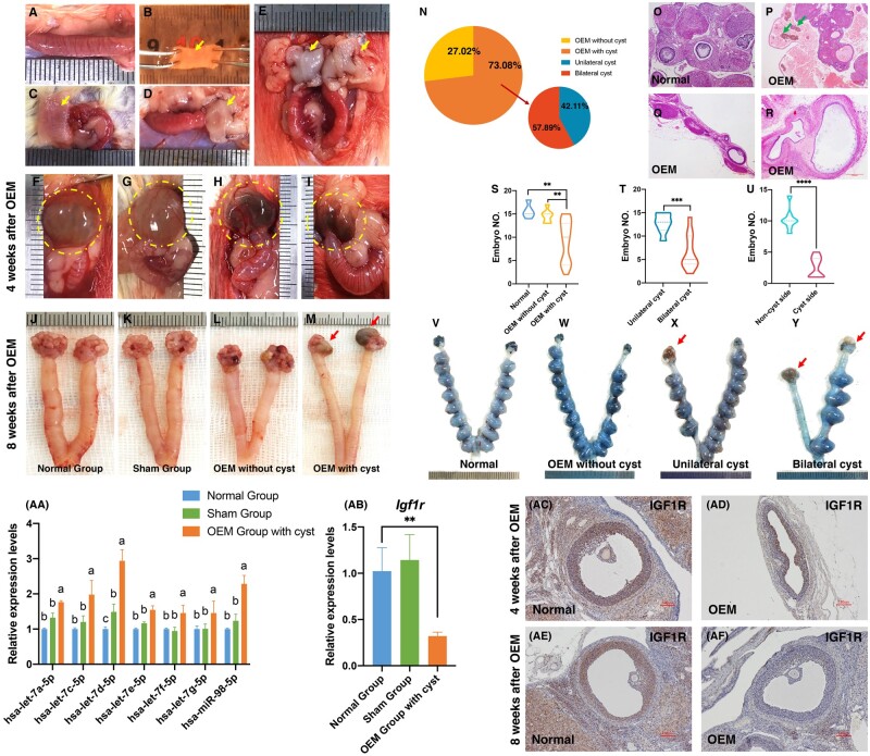
Study design, size, duration: This is a cross-sectional study aimed to elucidate the molecular mechanisms underlying the impact of OEM on follicular development. Initially, miRNA expression profiles in follicular fluid (FF) samples were sequenced from patients with infertility related to OEM (N = 3) and male factor (MF) infertility (N = 3), with the latter serving as the control group. Differentially expressed miRNAs were validated in additional samples from each group (N = 55 in OEM group and N = 45 in MF group) to confirm candidate miRNAs. The study also investigated indicators associated with GCs dysfunction in vitro on rat GCs. Subsequently, rat models of OEM were established through endometrial allogeneic transplantation, and fertility experiments were conducted to assess the let-7/IGF1R axis response to OEM in vivo. Patient samples were collected between May 2018 and April 2019, and the mechanistic study was conducted over the subsequent three years.

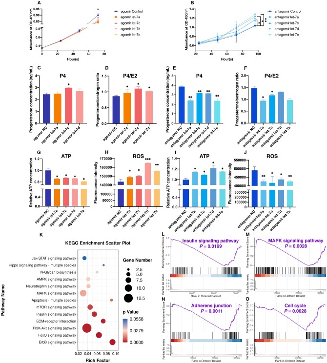
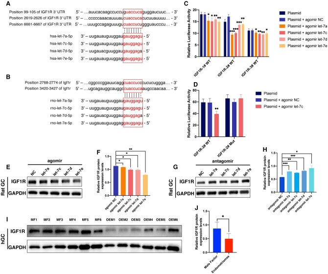
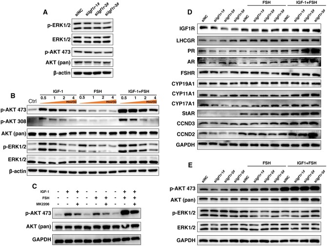
Participants/materials, setting, methods: FF and GC samples were obtained from infertile patients undergoing IVF treatment for OEM and MF related infertility. miRNA expression profiles in FF samples were analyzed using second-generation high-throughput sequencing technology, and candidate miRNAs were validated through quantitative PCR (qPCR). In the in vitro experiments conducted with rat GCs, cell proliferation was assessed using the CCK-8 assay, while steroid hormone concentrations were measured using chemiluminescence. ATP content was determined with an ATP assay kit, and levels of ROS were quantified using flow cytometry. A dual luciferase reporter gene assay was employed to identify the target gene of let-7 based on the construction of a IGF1R reporter gene plasmid using 293T cells. Western blotting was utilized to evaluate the expression of IGF1R in GCs, as well as its downstream proteins, and changes in signaling pathways following let-7 agomir/antagomir transfection and/or Igf1r silencing. In the in vivo OEM rat models, alterations in ovarian structure and cyst morphology were observed using hematoxylin and eosin staining. The expressions of let-7 and Igf1r in GCs were evaluated through qPCR, while variations in IGF1R expression were investigated with immunohistochemistry.

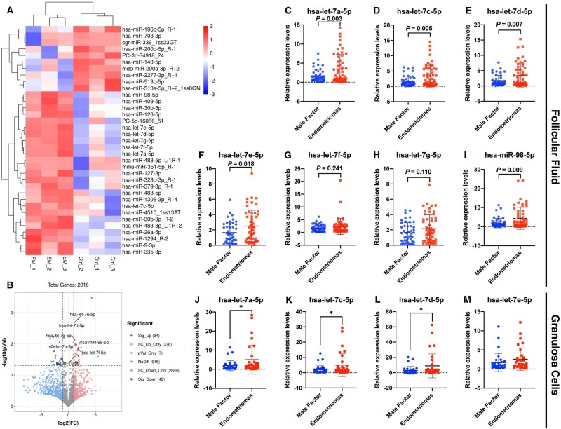
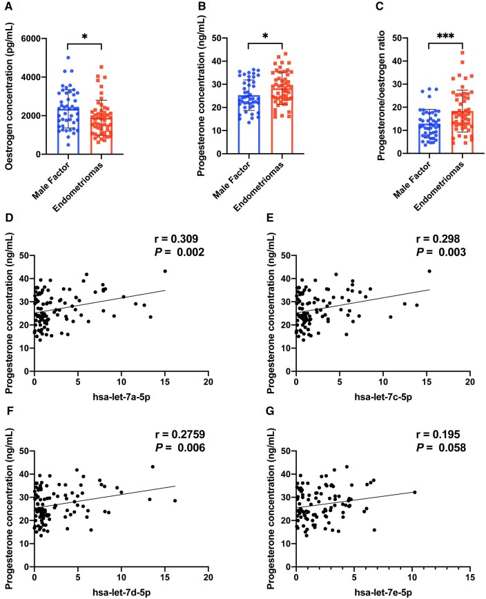
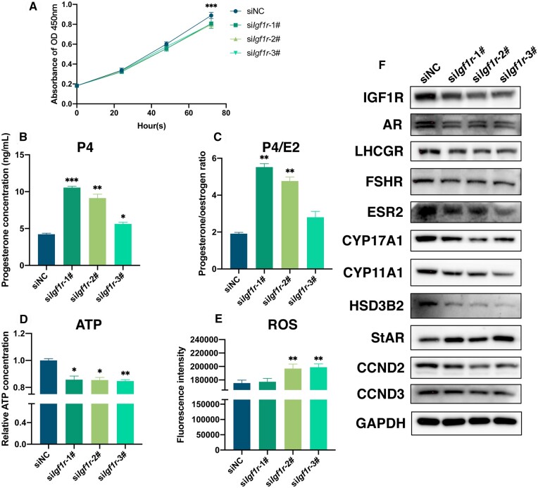
Main results and the role of chance: The cohort of patients with ovarian OEM in this study exhibited significantly decreased antral follicle counts, oocyte retrieval numbers, and normal fertilization rates compared to the control group with MF. The expression of the let-7 miRNA family was markedly upregulated in the FF and GCs of OEM patients. Transfection of rat GCs with let-7 agonists diminished the functions of GCs, including disrupted cell proliferation, mitochondrial oxidative phosphorylation, and steroid hormone secretion, while transfection of rat GCs with let-7 antagonists caused the opposite effects. Luciferase reporter gene experiments confirmed that let-7 complementarily bound to the 3'-untranslated regions of IGF1R. Stimulation of let-7 expression in rat GCs led to a significant decrease in IGF1R expression, while inhibition of let-7 increased IGF1R expression. The expression of IGF1R in the GCs of OEM patients was also significantly reduced compared to MF patients. Silencing of Igf1r led to the dysfunction of GCs, similar to the effects of let-7 agonization, as demonstrated by the downregulation of key proteins involved in cell proliferation (CCND2 and CCND3) and oestradiol synthesis, as well as an increase in progesterone synthesis (StAR), while implicating the PI3K-Akt and MAPK signaling pathways. The antagonistic effect of let-7 on GCs was ineffective when Igf1r was silenced. Conversely, the agonistic effect of let-7 on GCs could be reversed by stimulation with the IGF1R ligand IGF-1. These findings suggested that let-7 regulated the proliferation, differentiation, and ATP synthesis of GCs through targeting IGF1R. The OEM rat model demonstrated alterations in ovarian morphology and structure, along with reduced fertility. Let-7 expression was significantly upregulated in GCs of OEM rats compared to normal rats, while Igf1r and IGF1R expression in pre-ovulatory follicular GCs were notably downregulated, supporting the notion that elevated let-7 expression in the follicular microenvironment of OEM inhibited IGF1R, leading to abnormal GC function and impacting fertility at the molecular level.

Large scale data: N/A.

Limitations, reasons for caution: The synthesis and secretion mechanisms of steroid hormones are intricate and complex. Some enzymes that regulate oestrogen synthesis also play a role in progesterone synthesis. Moreover, certain receptors can respond to multiple hormone signals. Therefore, in this study, the expression patterns of key enzymes such as CYP17A, CYP11A1, HSD3B2, StAR, and receptors including AR, LHCGR, FSHR, ESR2, might be influenced by various factors and might not demonstrate complete consistency.

Wider implications of the findings: Future research will concentrate on investigating the potential impact of ovarian stromal cells on the external microenvironment of follicle growth. Additionally, screening for small molecule drugs that target let-7 and IGF1R actions can be conducted to intervene and modify the ovarian microenvironment, ultimately enhancing ovarian function.

Study funding/competing interest(s): This study received funding from the National Natural Science Foundation of China (grant number 82301851 to L.B.S., grant numbers U23A20403 and U20A20349 to S.Y.Z., and grant number 82371637 to Y.D.D.) and the Natural Science Foundation of Zhejiang Province (grant LTGY23H040010 to F.Z.). The authors have no conflicts of interest to declare.

Keywords: IGF1R; endometriosis; female infertility; follicular fluid; granulosa cells; let-7; ovarian endometriomas (OEM).


Conflict of interest statement

The authors have no conflicts of interest to declare.

Figures

Figure 1.
Figure 1.
miRNA sequencing of follicular fluid (FF) from patients with infertility associated with ovarian endometriomas (OEM) or male factor (MF), and validation of let-7 expression in FF and granulosa cells (GCs). (A) Cluster analysis of miRNA expression levels was shown, with the x-axis representing samples and the y-axis indicating the differentially expressed miRNAs (NMF = 3, NOEM = 3). (B) Volcano map analysis displayed differentially expressed miRNAs, with significantly upregulated miRNAs in red, downregulated miRNAs in blue, and non-significant miRNAs in gray. (C–I) Validation of hsa-let-7a-5p, hsa-let-7c-5p, hsa-let-7d-5p, hsa-let-7e-5p, hsa-let-7f-5p, hsa-let-7g-5p, and hsa-miR-98-5p in FF was conducted by expanding clinical samples through real-time quantitative PCR (qPCR) between the OEM and MF groups (NMF = 45, NOEM = 55). (J–M) Validation of hsa-let-7a-5p, hsa-let-7c-5p, hsa-let-7d-5p, and hsa-let-7e-5p in GCs was performed by expanding clinical samples through qPCR between the OEM and MF groups (NMF = 32, NOEM = 32). *P < 0.05.
Figure 2.
Figure 2.
Oestrogen and progesterone concentrations in follicular fluid (FF), and correlation analysis of let-7 expression and progesterone concentration. (A) Detection of oestrogen concentration in FF between the ovarian endometriomas (OEM) and male factor (MF) infertility groups. (B) Detection of progesterone concentration in FF between the OEM and MF groups. (C) Calculation of progesterone/oestrogen ratio in FF between the OEM and MF groups. (D) Correlation analysis of hsa-let-7a-5p expression and progesterone concentration. (E) Correlation analysis of hsa-let-7c-5p expression and progesterone concentration. (F) Correlation analysis of hsa-let-7d-5p expression and progesterone concentration. (G) Correlation analysis of hsa-let-7e-5p expression and progesterone concentration. NMF = 45, NOEM = 55, r, correlation coefficient, *P < 0.05, ***P < 0.001.
Figure 3.
Figure 3.
Effects of transfection of let-7 into rat primary granulosa cells (GCs). (A) Assessment of rat GCs proliferation within 72 h after transfection with let-7 agomir. (B) Assessment of rat GCs proliferation within 96 h after transfection with let-7 antagomir. (C) Measurement of progesterone concentration in rat GCs culture supernatant following let-7 agomir transfection. (D) Calculation of progesterone/oestrogen ratio in rat GCs culture supernatant post let-7 agomir transfection. (E) Evaluation of progesterone concentration in rat GCs culture supernatant after let-7 antagomir transfection. (F) Determination of progesterone/oestrogen ratio in rat GCs culture supernatant following let-7 antagomir transfection. (G) Adenosine triphosphate (ATP) detection in rat GCs post let-7 agomir transfection. (H) Reactive oxygen species (ROS) detection in rat GCs post let-7 agomir transfection via flow cytometry. (I) ATP detection in rat GCs post let-7 antagomir transfection. (J) ROS detection in rat GCs post let-7 antagomir transfection via flow cytometry. (K) Kyoto Encyclopedia of Genes and Genomes (KEGG) bubble chart illustrating the enrichment of signaling pathways post-transfection of let-7a agomir in rat GCs. Upregulated genes were depicted as red bars, while downregulated genes were represented by blue bars. (L–O) Gene set enrichment analysis (GSEA) demonstrating significant enrichment of signaling pathways post let-7a agomir transfection in rat GCs. *P < 0.05, **P < 0.01, ***P < 0.001.
Figure 4.
Figure 4.
Identification of IGF1R as the target gene of let-7. (A) The let-7 family bound to three complementary sites in the 3′-untranslated region of the human IGF1R gene. (B) The let-7 family bound to two complementary sites in the rat Igf1r gene. (C) Validation of the interaction between let-7 and IGF1R was performed through a dual luciferase reporter assay, confirming the presence of the three complementary binding sites. (D) Further validation of the interaction between hsa-let-7c-5p and the IGF1R-2# site was conducted using the dual luciferase reporter gene. (E) Detection of IGF1R expression after transfection of let-7 agomir into rat granulosa cells (GCs) through western blotting. (G) Detection of IGF1R expression after transfection of let-7 antagomir into rat GCs via western blotting. (I) Evaluation of IGF1R expression in human GCs from the ovarian endometriomas (OEM) and male factor (MF) groups through western blotting (NMF = 6, NOEM = 6). (F, H, and J) represent the quantitative analysis of (E, G, and I), respectively. *P < 0.05, **P < 0.01, ***P < 0.001.
Figure 5.
Figure 5.
Evaluation of granulosa cell (GC) function after silencing Igf1r in rat GCs. (A) Assessment of rat GCs proliferation within 72-h post-Igf1r silencing. (B) Measurement of progesterone concentration in rat GCs culture supernatant following siIgf1r transfection. (C) Calculation of progesterone/oestrogen ratio in rat GCs culture supernatant post-siIgf1r transfection. (D) Adenosine triphosphate detection in rat GCs post-Igf1r silencing. (E) Reactive oxygen species detection in rat GCs post-siIgf1r transfection via flow cytometry. (F) Western-blotting detection of the transfection efficiency of IGF1R post-Igf1r silencing, and the expressions of AR, LHCGR, FSHR, ESR2, CYP17A1, CYP11A1, HSD3B2, StAR, CCND2, and CCND3 with GAPDH as the reference protein. *P < 0.05, **P < 0.01, ***P < 0.001.
Figure 6.
Figure 6.
Detection of changes in the MAPK and PI3K-Akt signaling pathways after silencing Igf1r in rat granulosa cells (GCs). (A) Detection of p-ERK1/2, ERK1/2, p-AKT 473, and AKT (pan) expression following siIgf1r transfection. (B) Detection of p-AKT 473, p-AKT 308, AKT (pan), p-ERK1/2, and ERK1/2 expression after stimulating with IGF-1/FSH within 4 h. (C) Detection of p-AKT 473 and AKT (pan) expression after co-culturing with a PI3K-Akt signaling pathway inhibitor (MK2206) prior to IGF-1/FSH stimulation. (D) Detection of the expressions of IGF1R, LHCGR, PR, AR, FSHR, CYP19A1, CYP11A1, CYP17A1, StAR, CCND3, and CCND2 post-Igf1r silencing, and the reverse effects with IGF-1/FSH stimulation. (E) Detection of the expressions of p-AKT 473, AKT (pan), p-ERK1/2, and ERK1/2 after siIgf1r transfection, and the reverse effects induced by IGF-1/FSH.
Figure 7.
Figure 7.
let-7 causes dysfunction of granulosa cells (GCs) through targeting Igf1r. (A) Detection of the expressions of IGF1R, LHCGR, CYP19A1, CYP17A1, StAR, p-AKT 473, AKT (pan), p-ERK1/2, and ERK1/2 following let-7c antagomir transfection, and let-7c antagomir plus Igf1r silencing, demonstrating that the antagonistic effects of let-7 on GC function could be interrupted by Igf1r silencing. (B) Detection of the expression of the same group of proteins after transfection with let-7c agomir, followed by stimulation with IGF-1/FSH, indicating that the agonistic effect of let-7 on GC function could be reversed by IGF-1/FSH stimulation. (C) Measurement of progesterone concentration in rat GCs after let-7c antagomir transfection plus Igf1r silencing. (D) Calculation of progesterone/oestrogen ratio after let-7c antagomir transfection plus Igf1r silencing. (E) Adenosine triphosphate (ATP) detection after let-7c antagomir transfection plus Igf1r silencing. (F) ATP detection following transfection with let-7c agomir, and subsequent stimulation with IGF-1/FSH. (G) Detection of reactive oxygen species (ROS) in rat GCs via flow cytometry after transfection with let-7c antagomir in conjunction with Igf1r silencing. (H) ROS detection in rat GCs using flow cytometry after transfection with let-7c agomir, and subsequent stimulation with IGF-1/FSH. *P < 0.05, **P < 0.01, ***P < 0.001.
Figure 8.
Figure 8.
Establishment of the ovarian endometriomas (OEM) rat model and identification of the let-7/Igf1r axis in the follicular microenvironment. (A–E) Establishment of the OEM rat model through endometrial allogeneic transplantation. (F–I) Visualization of OEM cysts 4-week post-endometrium transplantation. (J–M) Visualization of OEM cysts 8-week post-endometrium transplantation. (N) Assessment of OEM cyst formation rate. (O–R) Histological examination of OEM cyst morphology using hematoxylin and eosin staining. (S) Comparison of embryo formation among normal rats, rats with and without OEM. (T) Comparison of embryo formation between rats with unilateral OEM cysts and rats with bilateral OEM cysts. (U) Comparison of embryo formation between the cystic and non-cystic sides in rats with unilateral OEM cysts. (V–Y) Illustration of embryo formation in normal rats (V), rats without OEM cysts (W), rats with unilateral OEM cysts (X), and rats with bilateral OEM cysts (Y). (Yellow arrows indicate fresh endometrium from donor rats, green arrows indicate the iron-containing heme in the fluid within the sac, and red arrows indicate OEM cysts.) (AA) Comparison of let-7 expression in granulosa cells (GCs) among rats in the normal group, sham group, and OEM group with ovarian cysts. Groups with different superscript letters above the columns indicate significant differences in the expression of let-7. (AB) Comparison of Igf1r expression in GCs among rats in the normal group, sham group, and OEM group with ovarian cysts. (AC–AF) Immunohistochemical analysis of IGF1R expression in pre-ovulation follicles of OEM cyst ovaries after 4 or 8 weeks of OEM model establishment. **P < 0.01, ***P < 0.001, ****P < 0.0001.
Figure 9.
Figure 9.
Schematic diagram of this study. Ovarian endometriomas induce the upregulation of let-7 expression in follicular fluid, resulting in dysfunction of granulosa cells through targeting of IGF1R.

References

    1. Alshehre SM, Narice BF, Fenwick MA, Metwally M. The impact of endometrioma on in vitro fertilisation/intra-cytoplasmic injection IVF/ICSI reproductive outcomes: a systematic review and meta-analysis. Arch Gynecol Obstet 2021;303:3–16. - PMC - PubMed
    1. Bagga S, Bracht J, Hunter S, Massirer K, Holtz J, Eachus R, Pasquinelli AE. Regulation by let-7 and lin-4 miRNAs results in target mRNA degradation. Cell 2005;122:553–563. - PubMed
    1. Baumgarten SC, Armouti M, Ko C, Stocco C. IGF1R expression in ovarian granulosa cells is essential for steroidogenesis, follicle survival, and fertility in female mice. Endocrinology 2017;158:2309–2318. - PMC - PubMed
    1. Bulun SE, Yilmaz BD, Sison C, Miyazaki K, Bernardi L, Liu S, Kohlmeier A, Yin P, Milad M, Wei J. Endometriosis. Endocr Rev 2019;40:1048–1079. - PMC - PubMed
    1. Busacca M, Vignali M. Ovarian endometriosis: from pathogenesis to surgical treatment. Curr Opin Obstet Gynecol 2003;15:321–326. - PubMed

MeSH terms