. 2024 Nov 9;15(11):810.
doi: 10.1038/s41419-024-07204-5.

Aurora B inhibitors promote RB hypophosphorylation and senescence independent of p53-dependent CDK2/4 inhibition

Affiliations

Aurora B inhibitors promote RB hypophosphorylation and senescence independent of p53-dependent CDK2/4 inhibition

Shivam Vora et al. Cell Death Dis. .

Abstract

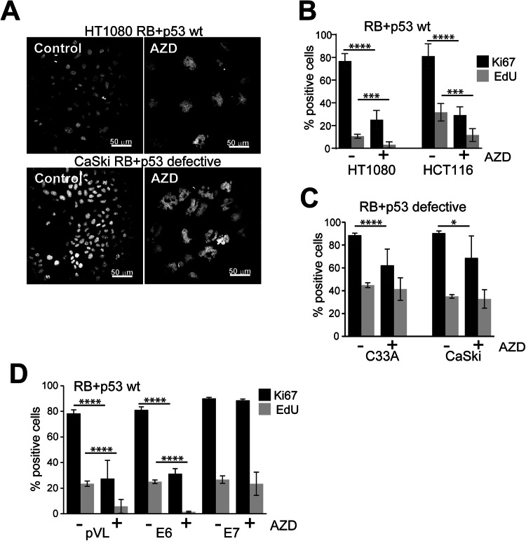
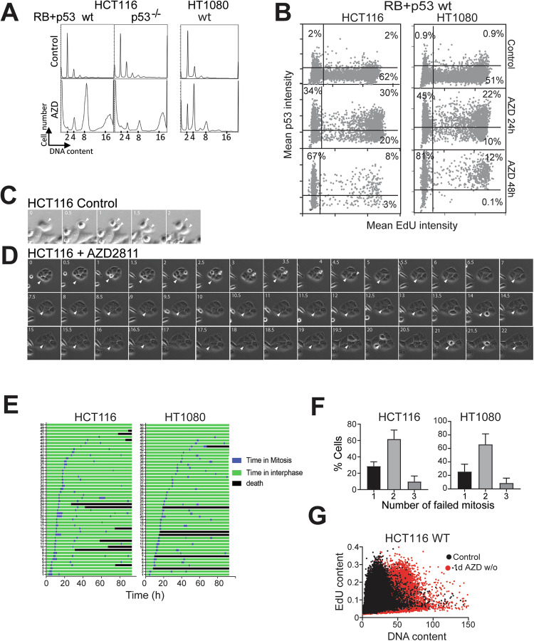
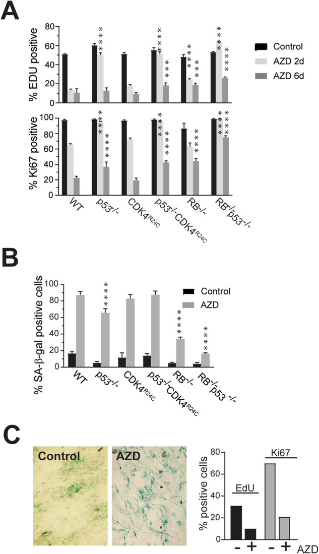
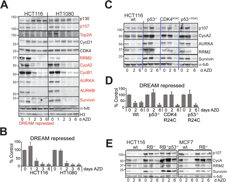
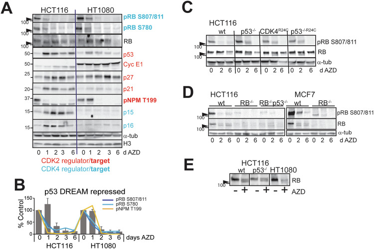
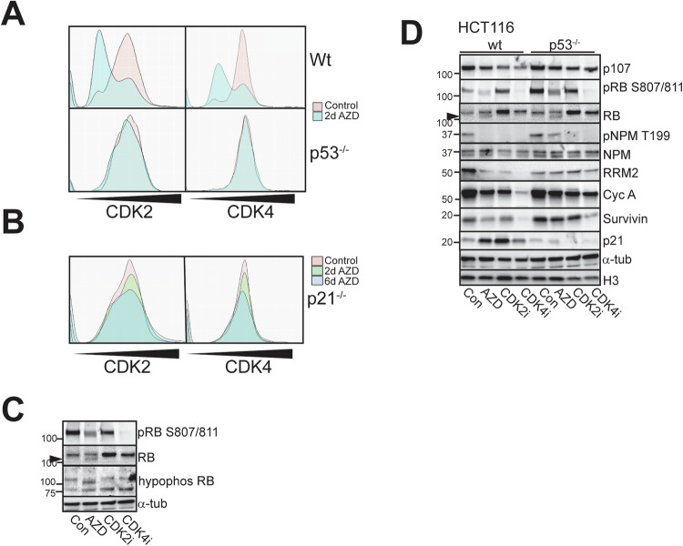
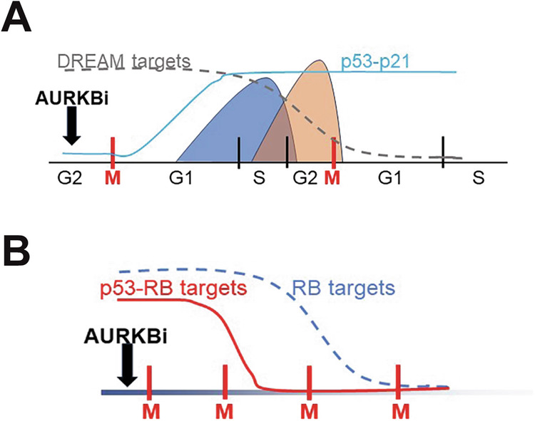
Aurora B kinase (AURKB) inhibitors have been trialled in a range of different tumour types but are not approved for any indication. Expression of the human papilloma virus (HPV) oncogenes and loss of retinoblastoma (RB) protein function has been reported to increase sensitivity to AURKB inhibitors but the mechanism of their contribution to sensitivity is poorly understood. Two commonly reported outcomes of AURKB inhibition are polyploidy and senescence, although their relationship is unclear. Here we have investigated the major cellular targets of the HPV E6 and E7, p53 and RB, to determine their contribution to AURKB inhibitor induced polyploidy and senescence. We demonstrate that polyploidy is a universal feature of AURKB inhibitor treatment in all cell types including normal primary cells, but the subsequent outcomes are controlled by RB and p53. We demonstrate that p53 by regulating p21 expression is required for an initial cell cycle arrest by inhibiting both CDK2 and CDK4 activity, but this arrest is only triggered after cells have undergone two failed mitosis and cytokinesis. However, cells can enter senescence in the absence of p53. RB is essential for AURKB inhibitor-induced senescence. AURKB inhibitor induces rapid hypophosphorylation of RB independent of inhibition of CDK2 or CDK4 kinases and p53. This work demonstrates that p53 activation determines the timing of senescence onset, but RB is indispensable for senescence.


Conflict of interest statement

JT, JS and JU are current or former employees and shareholders of AstraZeneca. Other authors declare no conflict of interest

Figures

Fig. 1
Fig. 1. Loss of RB and p53 bypasses AURKBi-induced cell cycle arrest.
A RB+p53 wild type HT1080 and RB+p53 defective CaSki cells were treated for 3 days with and without (control) 0.2 μM AZD2811 (AZD; AURKBi) then fixed and stained for DNA. Bars = 50 μm. B RB+p53 wild type HT1080 and HCT116 cells, C RB+p53 defective C33A and CaSki cells, and D immortalised human cervical keratinocytes expressing either empty vector (pLV), HPV E6 or E7 oncogenes, were treated for 6 days with 0.2 μM AZD2811, labelled for 2 h with EdU then stained for Ki67 and EdU incorporation. Cells were imaged using high content imaging and >1500 cells/well from at least three replicate wells were analysed. The percent positive staining is shown. Mean ± SD are shown. Data was collected from at least two independent experiments. Nonparametric t-test were used to compare control and treated data. The absence of comparison indicates no significant difference (***p > 0.001, ****p > 0.0001).
Fig. 2
Fig. 2. P53-induced cell cycle arrest occurs after 2 failed cytokinesis.
A RB+p53 wild type (wt) HCT116 and HT1080, and p53 deleted HCT116 cell lines were treated without (Control) or with 0.2 μM AZD2811 for 48 h then harvested for flow cytometry of DNA content. The data is representative of three independent experiments. B HCT116 and HT1080 cells either untreated (Control) or treated with AZD2811 for 24 or 48 h were labelled with EdU for 2 h then fixed and stained for EdU incorporation and p53, then >1000 cells/well in triplicate were analysed by high content image analysis. The data represents the pooled wells. The level of EdU and p53 staining in each cell is shown and percentage of cells in each quadrant shown. C, D HCT116 wild type cells were either untreated (Control) or treated with 0.2 μM AZD2811 then followed by time lapse microscopy, imaging every 30 min. A control mitosis and cytokinesis is shown (D white arrowheads) and two failed division in AZD2811-treated cells (D white arrow heads). E HCT116 and HT1080 cells were treated with 0.2 μM AZD2811 and followed by time lapse microscopy. Timelines for 50 cells in each cell line are shown, with the time in mitosis shown. All cells failed cytokinesis. F Analysis of time lapse imaging of AZD2811 treated HCT116 and HT1080 cells to quantify the number of mitoses each cell underwent over 80 h of treatment. Data are mean and SD for 100 cells were followed for each cell line from three independent experiments. G Cells from the control and cultures treated with AZD2811 for 1 day then drug washed out, were allowed to proliferate for a week. The resultant cultures were pulsed with EdU for 2 h to identify the proliferating population, and then fixed, stained and >1000 cells/well in triplicate were analysed by high content image analysis. The data represents the pooled wells. The EdU incorporation and DNA content for the control and AZD2811-treated cultures was overlaid.
Fig. 3
Fig. 3. Only RB is indispensable for AURKBi-induced senescence.
A HCT116 cells with indicated genotypes were treated with 0.2 μM AZD2811 for two or six days. Cells were labelled with EdU for 2 h then fixed. Cells were then stained for EdU and Ki67 and >1000 cells per well were quantitated by high content imaging. The data represent the mean and SD of four replicates from at least two independent experiments. B Cells treated as in (A) were fixed and stained for senescence associated β-galactosidase activity (SA-β-Gal). Quantitation of the SA-β-Gal positive cells in the indicated HCT116 genotypes. The data are the mean and SD from quantitating >four fields per cell line, each with >100 cells/field. Comparisons were to the equivalent treatment point in the WT control using two-way ANOVA with Tukey’s multiple comparison test. *** p < 0.001, ****p < 0.0001. C NFF cells were treated as in (A) then either stained for SA-β-Gal or labelled with EdU and Ki67 then >2000 cells analysed by high content imaging.
Fig. 4
Fig. 4. Loss of p53 only delays downregulation of critical cell cycle proteins.
A RB and p53 wild type HCT116 and HT1080 cells were treated for the indicated number of days with 0.2 μM AZD2811, then harvested and lysates immunoblotted for the indicated proteins. DREAM repressed cell cycle regulators (defined in [23]) are highlighted in red. This is representative of two independent experiments. B The levels of the DREAM repressed cell cycle regulators in (A) were quantified, expressed as percentage of control and combined. C The indicated HCT116 genotypes were treated for the indicated times with 0.2 μM AZD2811, harvested and lysates immunoblotted for the indicated proteins. The data are representative of replicate immunoblots. D The combined changes in cell cycle regulators from (C). E HCT116 wild type, RB deleted and RB+p53 deleted, MCF7 wild type and RB deleted lines were treated for the indicated times with 0.2 μM AZD2811 and immunoblotted for the indicated proteins. α-Tubulin was used as a loading control.
Fig. 5
Fig. 5. AURKBi downregulates CDK4 and CDK2 activity.
A HCT116 and HT1080 cells were treated for the indicated times with 0.2 μM AZD2811, then harvested and lysates immunoblotted for the indicated markers of CDK2 and CDK4 activity. The high mobility hypophosphorylated form of RB is indicated by the arrowhead in all panels. This is representative of replicate experiments. B The levels of phospho-RB and phospho-NPM were superimposed on the changes in the repressed cell cycle regulators (Fig. 4B). C The indicated HCT116 genotypes were treated for two or six days with 0.2 μM AZD2811 as in Fig. 4C and immunoblotted for RB and phospho-RB. D HCT116 wild type, RB deleted and RB and p53 deleted, MCF7 wild type and RB deleted lines were treated for the indicated times with AZD2811 and immunoblotted for the indicated proteins. The high mobility hypophosphorylated form of RB is indicated by the arrowhead. E HCT116 wild type and p53−/−, and HT1080 cells were treated without (Con) or with 0.2 μM AZD2811 for two days then harvested for immunoblotting for RB.
Fig. 6
Fig. 6. RB hypophosphorylation is independent of CDK4 and CDK2 activity.
A HCT116 wild type and p53−/− cells expressing both CDK2 and CDK4 biosensors were treated as indicated for two days then biosensor localisation was assessed by high content imaging and the activity calculated by determining the ratio of cytoplasmic to nuclear biosensor fluorescence. B HCT116 p21−/− cells expressing both biosensors were treated and analysed as in (A). For each experiment >2000 cells were analysed. The data are the average of triplicate experiments. C HCT116 cells, either untreated or treated with AZD2811 or 10 μM CDK2 or CDK4 selective inhibitors for two days were harvested and immunoblotted with indicated antibodies. D The indicated HCT116 genotypes were treated for two days with either 0.2 μM AZD2811 or 10 μM CDK2 or CDK4 inhibitors and cell lysates immunoblotted for the indicated markers.
Fig. 7
Fig. 7. Model for how p53 imposes a cell cycle arrest after two failed cytokinesis in AURKBi-treated cells.
A P53 is induced after the first failed cytokinesis but this is too late to block S phase progression or impose the DREAM repression of S/G2/M phase genes. However, these genes are cell cycle regulated at both mRNA and protein levels and will be downregulated after the second failed cytokinesis. B AURKBi imposed senescence by both increased p53 activity and hypophosphorylation of RB resulting repression of the overlapping RB and DREAM repressor gene expression. In the absence pf p53, hypophosphorylation of RB is sufficient to promote senescence albeit this is delayed.

References

    1. de Groot CO, Hsia JE, Anzola JV, Motamedi A, Yoon M, Wong YL, et al. A cell biologist’s field guide to Aurora kinase inhibitors. Front Oncol. 2015;5:285. - PMC - PubMed
    1. Vader G, Lens SM. The Aurora kinase family in cell division and cancer. Biochim Biophys Acta. 2008;1786:60–72. - PubMed
    1. Carmena M, Wheelock M, Funabiki H, Earnshaw WC. The chromosomal passenger complex (CPC): from easy rider to the godfather of mitosis. Nat Rev Mol Cell Biol. 2012;13:789–803. - PMC - PubMed
    1. Bavetsias V, Linardopoulos S. Aurora kinase inhibitors: current status and outlook. Front Oncol. 2015;5:278. - PMC - PubMed
    1. Marxer M, Ma HT, Man WY, Poon RY. p53 deficiency enhances mitotic arrest and slippage induced by pharmacological inhibition of Aurora kinases. Oncogene. 2014;33:3550–60. - PubMed

MeSH terms

LinkOut - more resources