. 2025 Jan;132(1):69-80.
doi: 10.1038/s41416-024-02892-4. Epub 2024 Nov 9.

VEGF-C propagates 'onward' colorectal cancer metastasis from liver to lung

Affiliations

VEGF-C propagates 'onward' colorectal cancer metastasis from liver to lung

Susanna Poghosyan et al. Br J Cancer. 2025 Jan.

Abstract

Background: The formation of lung metastasis as part of the progression of colon cancer is a poorly understood process. Theoretically, liver metastases could seed lung metastases.

Methods: To assess the contribution of the liver lymphatic vasculature to metastatic spread to the lungs, we generated murine liver-metastasis-derived organoids overexpressing vascular endothelial growth factor (VEGF)-C. The organoids were reimplanted into the mouse liver for tumour generation and onward metastasis.

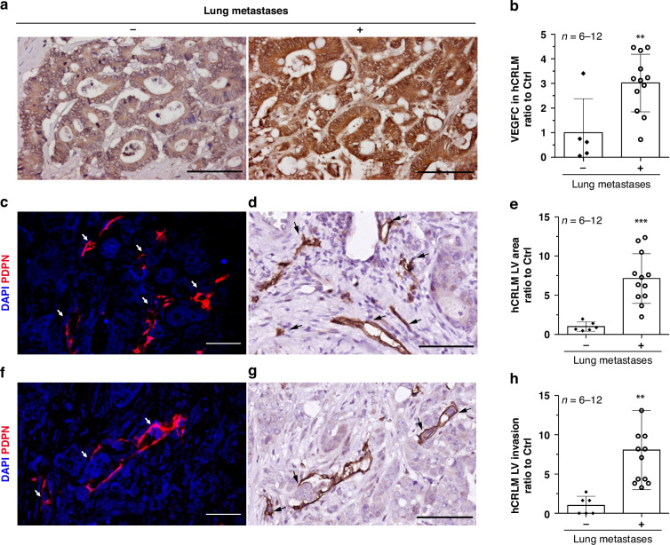
Results: Liver metastases from patients with concomitant lung metastases showed higher expression of VEGF-C, lymphatic vessel hyperplasia, and tumour cell invasion into lymphatic vessels when compared to those without lung metastases. Reimplantation of VEGF-C overexpressing organoids into the mouse liver showed that VEGF-C caused peritumoral lymphatic vessel hyperplasia, lymphatic tumour cell invasion, and lung metastasis formation. This change in metastatic organotropism was accompanied by reduced expression of WNT-driven adult stem cell markers, and increased expression of fetal stem cell markers and NOTCH pathway genes. Further NOTCH pathway inhibition with γ-secretase inhibitor (DAPT) in vivo results in a slight reduction in lung metastases and a decrease in lymphatic hyperplasia and invasion in VEGF-C-overexpressing tumours.

Conclusion: Collectively, these data indicate that VEGF-C can drive onward metastasis from the liver to the lung and suggest that targeting VEGF-C/NOTCH pathways may impair the progression of colorectal cancer.


Conflict of interest statement

Competing interests: The authors declare no competing interests. Ethics approval and consent to participate: All experiments with human tissues were approved by the Biobank Research Ethics Committee of the University Medical Centre Utrecht (Utrecht, the Netherlands). Written informed consent from the donors for the research use of tissue in this study was obtained prior to the acquisition of the specimen. The study work protocol (614-1-17) involving laboratory animals was approved by Utrecht University’s Animal Welfare Body, the Animal Ethics Committee and licensed by the Central Authority for Scientific Procedures on Animals (license numbers AVD115002016614, AVD11500202115055). All experiments were conducted in accordance with the Dutch Experiments on Animals Act, in line with European Directive 2010/63/EU and by licensed personnel. Consent for publication: Not applicable.

Figures

Fig. 1
Fig. 1. Increased VEGF-C expression and lymphatic hyperplasia in liver tumours of lung metastases-bearing CRC patients.
a Representative immunohistochemical analysis images of human CRC liver metastases (hCRLM) tumours for VEGF-C expression in patients with or without lung metastases. Scale bars: 100 µm. b Mean DAB area is quantified relative to the control (patients with no lung metastases) (n = 6–12). Representative high-resolution immunohistochemical analysis images of lymph vessels (LV) in hCRLM tumours. d, g H&E- or c, f DAPI-stained nuclei are shown in blue, and Podoplanin (PDPN) expressing lymphatic vessels and lymphatic vessels with invading tumour clusters are shown in d, g brown or c, f red. Scale bars: d, g for IHC images 50 µm, c, f for IF images 10 µm. e, h Mean DAB area is quantified relative to the control (patients with no lung metastases) (n = 6–12). Error bars are presented as SD from the arithmetic mean. Statistical significance on graphs is indicated p ≤ 0.01 by **p ≤ 0.001 by ***.
Fig. 2
Fig. 2. Lymphatic hyperplasia and increased lymphatic invasion in VEGF-Chigh liver tumours.
a Representative high-resolution immunohistochemical analysis images of lymph vessels (LV) in murine liver tumours. LVs are identified with LYVE1 lymphatic marker expression, where H&E- or DAPI-stained nuclei are shown in blue, and LYVE1-expressing lymphatic vessels are shown in brown or red. b DAB-positive tumour lymphatic vessel count was determined relative to the control (n = 5–6). c Representative high-resolution immunohistochemical analysis images of lymphatic invasion. LVs are identified with LYVE1 lymphatic marker expression, where H&E- or DAPI-stained nuclei are shown in blue, and LYVE1-expressing lymphatic vessels are shown in brown or red. Scale bars (a, c): for IHC images 25 µm, for IF images 10 µm. d DAB-positive tumour lymphatic vessels with invading tumour clusters were quantified relative to the control (n = 5–6). Error bars are presented as SD from the arithmetic mean. Statistical significance on graphs is indicated p ≤ 0.05 by *, p ≤ 0.01 by **, p ≤ 0.0001 by ****.
Fig. 3
Fig. 3. Increased lung metastasis in VEGF-Chigh organoid-implanted mice.
a Pictures of murine liver with tumour lesions in control and VEGF-Chigh mice. Dashed rectangle depicts the primary tumour implantation site. Scale bars: 5.3 mm. b Tumour tissue was identified using EpCam staining in immunohistochemical analysis. c in vivo tumour growth was monitored using bioluminescence imaging (cpm/cm2). d Bioluminescence imaging (cpm/cm2) of liver ex vivo from control and VEGF-Chigh mice. e Spread from the liver left implantation lobe to other liver lobes. EpCam expression was assessed using immunohistochemical analysis to determine the tumour area in the right, median and caudate lobes of liver from control and VEGF-Chigh mice n = 15–18. f Pictures of murine lungs in control and VEGF-Chigh mice. Scale bars: 3.2 mm. g Tumour lesions were identified using hematoxylin and eosin (H&E) staining. h Quantification of tumour area in lungs from control and VEGF-Chigh mice. n = 5–6. Scale bars: b, g full section images 1000 µm, b magnified liver sections 25 µm, g magnified lung sections 100 µm. Error bars are presented as SD from the arithmetic mean. Statistical significance on graphs is indicated p ≤ 0.001 by ***, p ≤ 0.0001 by ****.
Fig. 4
Fig. 4. Undifferentiated fetal-like phenotype in VEGF-Chigh liver tumours.
Heatmap of differentially expressed genes in control and VEGF-Chigh a liver tumours and b murine CRC liver organoids. cf RNA levels measured by RNA sequencing. Primary liver tumour tissue samples were sequenced in biological replicates; control (n = 5) and VEGF-Chigh (n = 3). g Immunohistochemical analysis of differentially expressed proteins in control and VEGF-Chigh liver tumours. Mean DAB area was quantified relative to the control (n = 5–6). Scale bars: 50 µm. Error bars are presented as SD from the arithmetic mean. Statistical significance on graphs is indicated p ≤ 0.05 by *, p ≤ 0.001 by ***, p ≤ 0.0001 by ****.
Fig. 5
Fig. 5. Increased NOTCH1 expression in VEGF-Chigh liver tumours and invading lymphatic vessels.
Multiplex immunohistochemical analysis of control and VEGF-Chigh liver tumours (n = 4–6). a DAPI-stained nuclei are shown in blue, and NOTCH1 expression is shown in green. Scale bars: 25 µm. b Representative images of increased NOTCH1 expression in VEGF-Chigh liver tumours and lymphatic vessels with invading tumour clusters. DAPI-stained nuclei are shown in blue, and NOTCH1 expression is shown in green, LYVE1 expression in lymphatic vessels is shown in red. White arrows indicate lymphatic vessel-invading tumour clusters. Scale bars: 10 µm.
Fig. 6
Fig. 6. Reduced viability after NOTCH inhibition in VEGF-Chigh murine CRC organoids.
a Representative immunofluorescence analysis images of control and VEGF-Chigh murine CRC organoids untreated or treated with NOTCH inhibitor (DAPT). DAPI-stained nuclei are shown in blue, and Caspase-3 expression is shown in red. b Mean fluorescence intensity was quantified relative to the control (n = 8–15). Scale bars: 10 µm. Error bars are presented as SD from the arithmetic mean. Statistical significance on the graph is indicated p ≤ 0.05 by *, p ≤ 0.0001 by ****.
Fig. 7
Fig. 7. Increased NOTCH1 expression in CRC liver tumours and invading lymphatic vessels of lung metastases-bearing CRC patients.
a Representative immunohistochemical analysis images of hCRLM tumours for NOTCH1 expression in patients with or without lung metastases. Scale bars: 15 µm. Mean DAB area was quantified relative to the control (patients with no lung metastases) (n = 6–12). Representative multiplex high-resolution immunohistochemical analysis images of hCRLM tumours from patients b with lung metastases and c without lung metastases. DAPI-stained nuclei are shown in blue, and Podoplanin (PDPN) expressing lymphatic vessels are shown in red, NOTCH1 expression is shown in green. White arrows indicate lymphatic vessel-invading tumour clusters Scale bars: 18 µm.
Fig. 8
Fig. 8. Reduced lung metastasis and lymphatic hyperplasia in NOTCH-inhibited VEGF-Chighmice.
a Lung tumour lesions were identified using hematoxylin and eosin (H&E) staining. Quantification of tumour area in lungs from control and VEGF-Chigh mice with or without DAPT treatment (n = 8–10). b Immunohistochemical analysis of DAPT-treated or untreated tumours for LYVE1 lymphatic marker expression. DAB-positive tumour lymphatic vessel count was determined relative to the untreated control. c Immunohistochemical analysis of DAPT-treated or untreated tumours for lymphatic invasion. DAB-positive tumour lymphatic vessels with invading tumour clusters was determined relative to the untreated control. (n = 3). Error bars are presented as SD from the arithmetic mean. Statistical significance on the graph is indicated p ≤ 0.01 by **.
Fig. 9
Fig. 9
Schematic overview of CRC metastatic routes.

References

    1. Engstrand J, Nilsson H, Strömberg C, Jonas E, Freedman J. Colorectal cancer liver metastases—a population-based study on incidence, management and survival. BMC Cancer. 2018;18:78. - PMC - PubMed
    1. Hackl C, Neumann P, Gerken M, Loss M, Klinkhammer-Schalke M, Schlitt HJ. Treatment of colorectal liver metastases in Germany: a ten-year population-based analysis of 5772 cases of primary colorectal adenocarcinoma. BMC Cancer. 2014;14:810. - PMC - PubMed
    1. Punt CJA, Koopman M, Vermeulen L. From tumour heterogeneity to advances in precision treatment of colorectal cancer. Nat Rev Clin Oncol. 2016;14:235–46. - PubMed
    1. Stewart CL, Warner S, Ito K, Raoof M, Wu GX, Kessler J, et al. Cytoreduction for colorectal metastases: liver, lung, peritoneum, lymph nodes, bone, brain. When does it palliate, prolong survival, potential cure?. Curr Probl Surg. 2018;55:330–79. - PMC - PubMed
    1. Petrowsky H, Fritsch R, Guckenberger M, De Oliveira ML, Dutkowski P, Clavien P-A. Modern therapeutic approaches for the treatment of malignant liver tumours. Nat Rev Gastroenterol Hepatol. 2020;17:755–72. - PubMed

MeSH terms

Substances

LinkOut - more resources