. 2024 Nov 9;15(1):411.
doi: 10.1186/s13287-024-04020-8.

Post-symptomatic administration of hMSCs exerts therapeutic effects in SCA2 mice

Affiliations

Post-symptomatic administration of hMSCs exerts therapeutic effects in SCA2 mice

Sehwan Kim et al. Stem Cell Res Ther. .

Abstract

Background: Defects in the ataxin-2 (ATXN-2) protein and CAG trinucleotide repeat expansion in its coding gene, Atxn-2, cause the neurodegenerative disorder spinocerebellar ataxia type 2 (SCA2). While clinical studies suggest potential benefits of human-derived mesenchymal stem cells (hMSCs) for treating various ataxias, the exact mechanisms underlying their therapeutic effects and interaction with host tissue to stimulate neurotrophin expression remain unclear specifically in the context of SCA2.

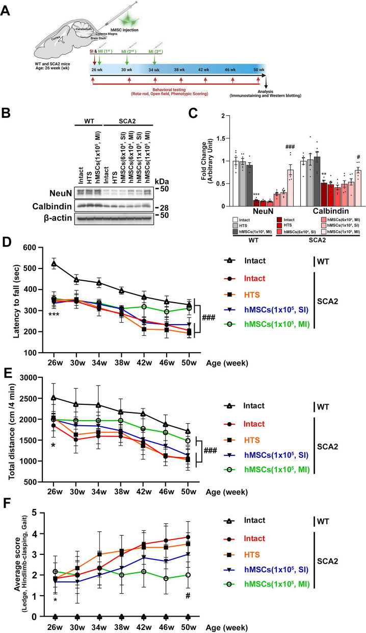
Methods: Human bone marrow-derived MSCs (hMSCs) were injected into the cisterna magna of 26-week-old wild-type and SCA2 mice. Mice were assessed for impaired motor coordination using the accelerating rotarod, open field test, and composite phenotype scoring. At 50 weeks, the cerebellum vermis was harvested for protein assessment and immunohistochemical analysis.

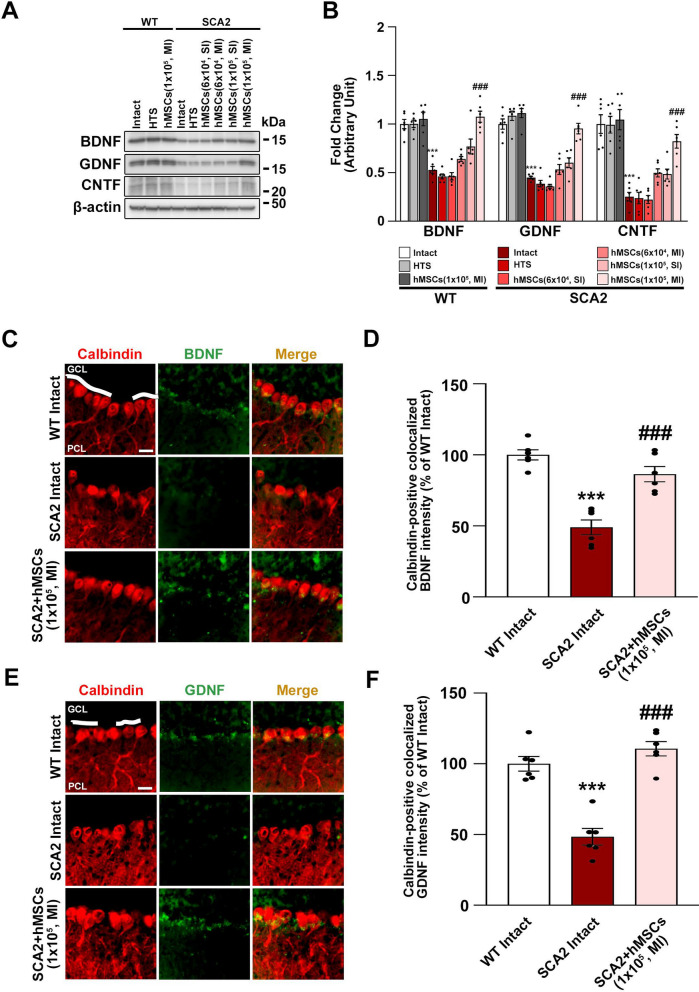
Results: Significant loss of NeuN and calbindin was observed in 25-week-old SCA2 mice. However, after receiving multiple injections of hMSCs starting at 26 weeks of age, these mice exhibited a significant improvement in abnormal motor performance and a protective effect on Purkinje cells. This beneficial effect persisted until the mice reached 50 weeks of age, at which point they were sacrificed to study further mechanistic events triggered by the administration of hMSCs. Calbindin-positive cells in the Purkinje cell layer expressed bone-derived neurotrophic factor after hMSC administration, contributing to the protection of cerebellar neurons from cell death.

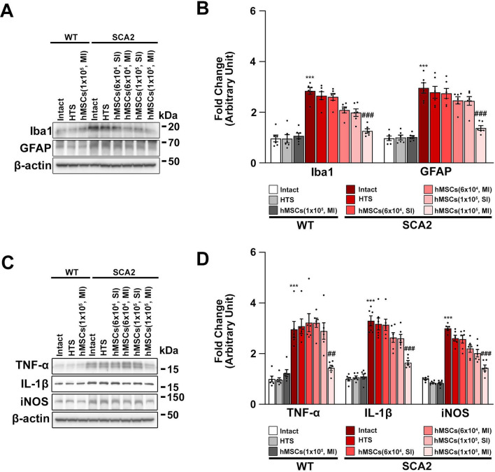
Conclusion: In conclusion, repeated administration of hMSCs shows promise in alleviating SCA2 symptoms by preserving Purkinje cells, improving neurotrophic support, and reducing inflammation, ultimately leading to the preservation of locomotor function in SCA2 mice.

Keywords: Follistatin-like 1; Mesenchymal stem cells; Neuroinflammation; Neurotrophic factor; Purkinje cells; Spinocerebellar ataxia type 2.


Conflict of interest statement

The authors declare no competing financial and non-financial interests.

Figures

Fig. 1
Fig. 1
Beneficial effects of human mesenchymal stem cells (hMSCs) on behavioral impairment and neuronal damage in spinocerebellar ataxia 2 (SCA2) mice. A Experiment schematic of hMSC treatment in wild-type (WT) and SCA2 mice. The figure was created using Biorender.com (Agreement number: XL26IE3A0R). hMSC treatment was administered once (single injection; SI) or thrice (multiple injections; MI) at 4-week intervals in 26-week-old WT and SCA2 mice. B, C Western blot analysis of neuronal nuclear protein (NeuN) and calbindin in the cerebellum after 24 weeks of hMSC treatment (50-week-old mice). **p < 0.01 and ***p < 0.001 vs. untreated WT mice; #p < 0.05 and ###p < 0.001 vs. treated SCA2 mice (two-way analysis of variance [ANOVA] with Tukey’s post-hoc analysis; n = 6 for each group). Full-length blots are presented in Supplementary Fig. 1 and Fig. 1B. D A behavioral test for motor coordination impairment was performed using the rotarod test in WT and SCA2 mice treated with hMSCs at 4-week intervals from 26 to 50 weeks of age. ***p < 0.001 vs. untreated WT mice; ###p < 0.001 vs. untreated SCA2 mice (one-way ANOVA with Tukey’s post hoc analysis; n = 6 for each group). E A behavioral test for locomotor activity was performed using the open-field test in WT and SCA2 mice at 4-week intervals after hMSC treatment. *p < 0.05 vs. untreated WT mice; ###p < 0.001 vs. untreated SCA2 mice (one-way ANOVA with Tukey’s post hoc analysis; n = 6 for each group). F A simple composite phenotype scoring system was used to evaluate WT and SCA2 mice at 4-week intervals after hMSC treatment. *p < 0.05 vs. untreated WT mice; #p < 0.05 vs. untreated SCA2 mice (one-way ANOVA with Tukey’s post-hoc analysis; Kruskal–Wallis test for average score; n = 6 for each group)
Fig. 2
Fig. 2
Treatment with hMSCs upregulates the protein levels of neurotrophic factors in the cerebellum of SCA2 mice. A, B Western blot analysis of brain-derived neurotrophic factor (BDNF), glial cell-derived neurotrophic factor (GDNF), and ciliary neurotrophic factor (CNTF) in the cerebellum after 24 weeks of hMSC administration (50-week-old mice). ***p < 0.001 vs. untreated WT mice; ###p < 0.001 vs. untreated SCA2 mice (two-way ANOVA with Tukey’s post-hoc analysis; n = 6 for each group). Full-length blots are presented in Supplementary Fig. 5 and Fig. 2A. C Double immunofluorescence staining was performed for calbindin and BDNF in the cerebellum of 50-week-old WT, SCA2, and SCA2 + hMSCs (MI, 1 × 105 cells)-treated mice. The scale bar represents 20 μm. A dashed line is used to differentiate between the granule cell layer and the Purkinje cell layer. D The fluorescence intensity for the quantitative co-localization of calbindin with BDNF and GDNF was measured. ***p < 0.001 vs. untreated WT mice; ###p < 0.001 vs. untreated SCA2 mice (one-way ANOVA with Tukey’s post-hoc analysis). E Double immunofluorescence staining was performed for calbindin and GDNF in the cerebellum of 50-week-old WT, SCA2, and SCA2 + hMSCs (MI, 1 × 105 cells)-treated mice. The scale bar represents 20 μm. A dashed line is used to differentiate between the granule cell layer and the Purkinje cell layer. F The fluorescence intensity for the quantitative co-localization of calbindin with GDNF was measured. ***p < 0.001 vs. untreated WT mice; ###p < 0.001 vs. untreated SCA2 mice (one-way ANOVA with Tukey’s post-hoc analysis)
Fig. 3
Fig. 3
Treatment with hMSCs downregulates the protein levels of glial-mediated neurotoxic cytokines in the cerebellum of SCA2 mice. A, B Western blot analysis of ionized calcium-binding adaptor molecule 1 (Iba1) and glial fibrillary acidic protein (GFAP) in the cerebellum after 24 weeks of hMSC administration (50-week-old mice). ***p < 0.001 vs. untreated WT mice; ###p < 0.001 vs. untreated SCA2 mice (two-way ANOVA with Tukey’s post hoc analysis; n = 6 for each group). Full-length blots are presented in supplymentary Fig. 5: Fig. 3A. C, D Western blot analysis of tumor necrosis factor-alpha (TNF-α), interleukin-1 beta (IL-1β), and inducible nitric oxide synthase (iNOS) in the cerebellum after 24 weeks of hMSC administration (50-week-old mice). ***p < 0.001 vs. untreated WT mice; ##p < 0.01 and ###p < 0.001 vs. untreated SCA2 mice (two-way ANOVA with Tukey’s post hoc analysis; n = 6 for each group). Full-length blots are presented in supplymentary Fig. 5: Fig. 3C
Fig. 4
Fig. 4
hMSC treatment preserves the protein levels of follistatin-like 1 (FSTL1) and transforming growth factor beta 1 (TGF-β1) in the cerebellum of SCA2 mice. A, B Western blot analysis of FSTL1 and TGF-β1 in cerebellum tissues obtained from 10-, 25-, and 50-week-old WT and SCA2 mice. **p < 0.001 and ***p < 0.001 vs. 10-week-old WT mice (two-way ANOVA with Tukey’s post hoc analysis; n = 6 for each group). Full-length blots are presented in supplementary Fig. 5; Fig. 4A. C Double immunofluorescence staining was performed for calbindin and FSTL1 as well as calbindin and TGF-β1 in the cerebellum of 50-week-old WT and SCA2 mice. The scale bar represents 20 μm. The dashed line differentiates between the granule and Purkinje cell layers. D The fluorescence intensity for the quantitative co-localization of calbindin with FSTL1 and TGF-β1 was measured. ***p < 0.001 vs. untreated WT mice (t-test with Tukey’s post-hoc analysis). E Double immunofluorescence staining was performed for NeuN and FSTL1 as well as NeuN and TGF-β1 in the cerebellum of 50-week-old WT and SCA2 mice. The scale bar represents 20 μm. The dashed line differentiates between the granule and Purkinje cell layers. F The fluorescence intensity for the quantitative co-localization of NeuN with FSTL1 and TGF-β1 was measured. ***p < 0.001 vs. untreated WT mice (t-test with Tukey’s post-hoc analysis). G, H Western blot analysis of FSTL1 and TGF-β1 in the cerebellum after 24 weeks of hMSC administration (50-week-old mice). ***p < 0.001 vs. untreated WT mice; ###p < 0.001 vs. untreated SCA2 mice (two-way ANOVA with Tukey’s post hoc analysis; n = 6 for each group). Full-length blots are presented in supplymentary Fig. 5: Fig. 4G. I Schematic representation of the effects of hMSCs in the SCA2 model. hMSC administration upregulates the expression of FSTL1, which in turn reduces inflammation and is accompanied by behavioral and physiological improvements in the SCA2 phenotype. The figure was created using BioRender.com (Agreement number: BG25IEMZTA)

Similar articles

References

    1. Schols L, Bauer P, Schmidt T, Schulte T, Riess O. Autosomal dominant cerebellar ataxias: clinical features, genetics, and pathogenesis. Lancet Neurol. 2004;3:291–304. - PubMed
    1. Auburger GW. Spinocerebellar ataxia type 2. Handb Clin Neurol. 2012;103:423–36. - PubMed
    1. Seidel K, Siswanto S, Brunt ER, den Dunnen W, Korf HW, Rub U. Brain pathology of spinocerebellar ataxias. Acta Neuropathol. 2012;124:1–21. - PubMed
    1. Sinha KK, Worth PF, Jha DK, Sinha S, Stinton VJ, Davis MB, Wood NW, Sweeney MG, Bhatia KP. Autosomal dominant cerebellar ataxia: SCA2 is the most frequent mutation in eastern India. J Neurol Neurosurg Psychiatry. 2004;75:448–52. - PMC - PubMed
    1. Lo RY, Figueroa KP, Pulst SM, Lin CY, Perlman S, Wilmot G, Gomez CM, Schmahmann J, Paulson H, Shakkottai VG, Ying SH, Zesiewicz T, Bushara K, Geschwind M, Xia G, Subramony SH, Ashizawa T, Kuo SH. Vascular risk factors and clinical progression in spinocerebellar ataxias. Tremor Other Hyperkinet Mov. 2015;5:287. - PMC - PubMed

MeSH terms

LinkOut - more resources