Case Reports
. 2024 Nov 9;24(1):436.
doi: 10.1186/s12883-024-03945-0.

Transient biopsy-proven progressive multifocal leukoencephalopathy-immune reconstitution inflammatory syndrome (PML-IRIS) in an elderly woman without known immunodeficiency: a case report

Affiliations
Case Reports

Transient biopsy-proven progressive multifocal leukoencephalopathy-immune reconstitution inflammatory syndrome (PML-IRIS) in an elderly woman without known immunodeficiency: a case report

Susanne Gaarden Ingebrigtsen et al. BMC Neurol. .

Abstract

Background: Progressive multifocal leukoencephalopathy (PML) is a severe opportunistic brain disease caused by lytic JC polyomavirus (JCPyV) replication in oligodendrocytes. Although JCPyV infection is common in the general population, PML almost exclusively occurs in patients immunocompromised due to untreated HIV/AIDS, haematological malignancies, primary immunodeficiencies, solid organ transplantation, or immunomodulatory treatment of autoimmune diseases. There is no effective antiviral treatment, and recovery depends on immune reconstitution. Paradoxically, initiation of antiretroviral therapy for HIV/AIDS or interruption of immunomodulating treatment can worsen the clinical manifestations due to immune reconstitution inflammatory syndrome (IRIS). Here, we report an unusual case of spontaneous IRIS in a 76-year-old immunocompetent woman, unmasking PML and leading to unexpected recovery.

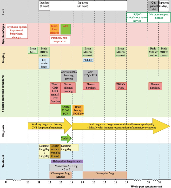
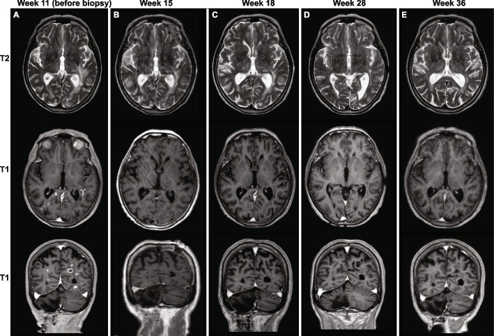
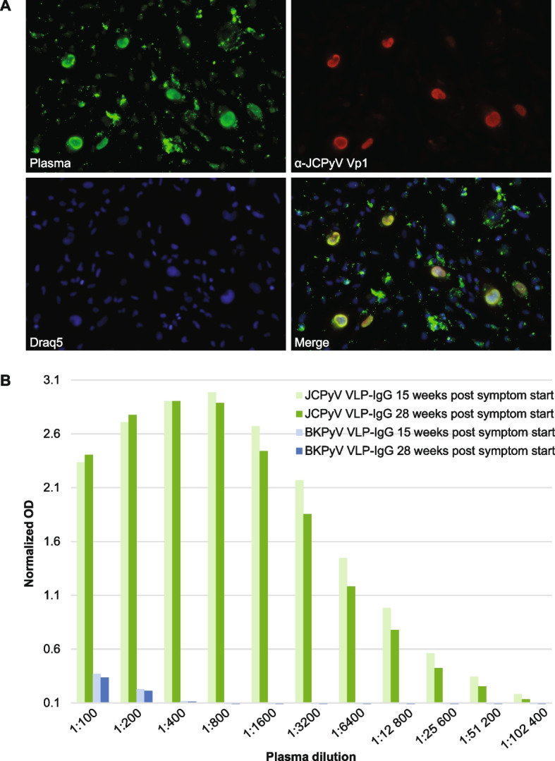
Case presentation: The patient was admitted to the hospital due to psychosis, speech impairment, and behavioral changes over the last three months. She had previously been healthy, except for a cerebellar stroke secondary to paroxysmal atrial fibrillation. Magnetic resonance imaging (MRI) revealed multiple contrast-enhancing white matter lesions suspicious of cancer metastases. Due to suspicion of edema, dexamethasone was administered, and the patient was released while waiting for a stereotactic brain biopsy. Eight days later, she suffered tonic seizures and was readmitted. Intravenous levetiracetam gave rapid effect, but the patient was paranoid and non-cooperative, and dexamethasone was unintentionally discontinued. Ten days later, the brain biopsy revealed demyelination, abundant perivascular T cells, macrophages, and scattered JCPyV-infected oligodendrocytes, rendering the diagnosis of PML-IRIS. The cerebrospinal fluid contained low amounts of JCPyV-DNA, and plasma contained high levels of anti-JCPyV immunoglobulin G. Despite extensive immunological testing, no evidence of immunodeficiency was found. The patient gradually recovered clinically and radiologically. More than 19 months after diagnosis, the patient has only a slight impairment in language and behavior.

Conclusions: An apparently immunocompetent elderly person developed clinically symptomatic PML, which spontaneously resolved with symptoms and signs of IRIS. The atypical MRI lesions with contrast enhancement and the lack of known immunological risk factors for PML delayed the diagnosis, eventually proved by biopsy. PML and PML-IRIS should be considered in the differential diagnosis of patients presenting CNS symptoms and focal lesions with contrast enhancement on MRI.

Keywords: Brain biopsy; CD8 + T cells; Case report; Flow cytometric immunophenotyping; Immune reconstitution inflammatory syndrome; JC polyomavirus; JCPyV antibody; MRI; Macrophages; Progressive multifocal leukoencephalopathy.


Conflict of interest statement

The authors declare no competing interests.

Figures

Fig. 1
Fig. 1
Timeline of symptoms and signs, selected diagnostic procedures, and treatment. On the X-axis, time is shown as weeks after the symptoms started
Fig. 2
Fig. 2
Chronological changes in brain magnetic resonance imaging (MRI). T2-flair on the top row and T1-flair on the next two rows. A 11 weeks after the symptoms started, i.e., 18 days before the biopsy, patchy T2 hyperintensities in the white matter are seen. The lesions are periventricular, mostly around the occipital- and temporal horns, with a left-sided predominance. There are also some subcortical lesions, nodular contrast enhancement in smaller lesions, and ring-like patterns in the more extensive lesions. The lesions are partly confluent with surrounding edema. B 15 weeks after the symptom started, i.e., 10 days after the biopsy, there is still some edema but almost complete regression of the contrast enhancement in the lesions. The patient was moving slightly. C 18 weeks after symptoms started, there were no or only minimal changes from the previous examination. D 28 weeks after symptoms started, there were no or only minimal changes from the previous examination. E 36 weeks after symptoms started, there were no or only minimal changes from the previous examination
Fig. 3
Fig. 3
Staining and immunohistochemistry of the brain biopsy. A Luxol fast blue staining shows demyelination in the border of the grey and white matter junction. The insert shows macrophages containing myelin. B Hematoxylin and Eosin staining showing the presence of extensive inflammatory infiltrates with perivascular lymphoid cuffing (white asterisk), foamy macrophages (white arrow), and reactive astrocytes (black arrow). In the insert, an oligodendrocyte with enlarged nuclei is seen (black asterisk). C CD3 and CD20 immunostaining showing T-cells (in brown) and B-cells (in red), respectively. More T than B-cells are present. D CD68 immunostaining shows plenty of macrophages. E ki67 immunostaining shows strong staining of enlarged oligodendrocytes and other cells. F p53 immunostaining shows strong staining of enlarged oligodendrocytes and other cells. G JCPyV large T-antigen immunostaining, using the cross-reacting SV40 antibody Pab416, shows scattered enlarged oligodendrocytes with nuclear staining. H JCPyV Vp1 immunostaining (ab34756) shows grainy cytoplasmic staining. Magnification for panels A, C, D, E, F, and G: overview 5x, insert 20x. Magnification for panel B: overview 10x, insert 40x, and for panel H: 20x
Fig. 4
Fig. 4
Serology JCPyV and BKPyV. A SVG-A cells were infected by JCPyV and fixed by methanol before immunofluorescence staining was performed using a combination of patient plasma from 15 weeks post symptom start and the mouse monoclonal anti-JCPyV Vp1 antibody (ab34756). Alexa 488-labelled goat-anti human IgG (green) and Alexa 568-labelled goat anti-mouse IgG (red) were used as secondary antibodies. Nuclei were labelled by Draq5 (blue). The plasma and the specific JCPyV Vp1 antibody show overlapping nuclear staining, suggesting that the patient has developed JCPyV Vp1 IgG. B JCPyV- and BKPyV-specific IgG using Vp1 virus-like particle (VLP)-based ELISAs. The two available serum samples were tested in several dilutions, and the result was normalized to a laboratory reference serum. The cut-off of the assay is 0.1. The plasma contained very high levels of JCPyV Vp1 IgG, as it could be diluted more than 100,000 times and still be positive. In contrast, it only contained low levels of BKPyV Vp1 IgG, which could only be detected with dilutions less than 800

References

    1. Hou J, Major EO. Progressive multifocal leukoencephalopathy: JC virus induced demyelination in the immune compromised host. J Neurovirol. 2000;6(Suppl 2):S98–100. - PubMed
    1. Gheuens S, Wuthrich C, Koralnik IJ. Progressive multifocal leukoencephalopathy: why gray and white matter. Annu Rev Pathol. 2013;8:189–215. 10.1146/annurev-pathol-020712-164018. - PubMed
    1. Atkinson AL, Atwood WJ. Fifty Years of JC Polyomavirus: A Brief Overview and Remaining Questions. Viruses. 2020;12:9. 10.3390/v12090969. - PMC - PubMed
    1. Knowles WA. Discovery and epidemiology of the human polyomaviruses BK virus (BKV) and JC virus (JCV). Adv Exp Med Biol. 2006;577:19–45. 10.1007/0-387-32957-9_2. - PubMed
    1. Egli A, Infanti L, Dumoulin A, Buser A, Samaridis J, Stebler C, et al. Prevalence of Polyomavirus BK and JC Infection and Replication in 400 Healthy Blood Donors. J Infect Dis. 2009;199:837–46. 10.1086/597126. - PubMed

Publication types

MeSH terms

LinkOut - more resources