

Endogenous ZAP is associated with altered Zika virus infection phenotype
- PMID: 39522048
- PMCID: PMC11549788
- DOI: 10.1186/s12985-024-02557-x
Endogenous ZAP is associated with altered Zika virus infection phenotype
Abstract
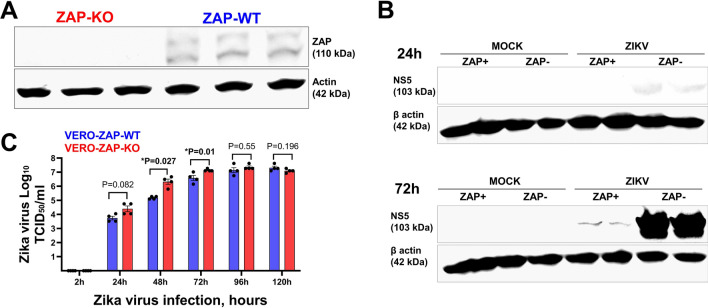
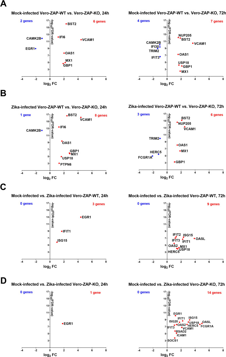
The zinc finger antiviral protein 1 (ZAP) has broad antiviral activity. ZAP is an interferon (IFN)-stimulated gene, which itself may enhance type I IFN antiviral response. In a previous study, Zika virus was identified as ZAP-resistant and not sensitive to ZAP antiviral activity. Here, we found that ZAP was associated with the inhibition of Zika virus in Vero cells, in the absence of a robust type I IFN system because Vero cells are deficient for IFN-alpha and -beta. Also, quantitative RNA-seq data indicated that endogenous ZAP is associated with altered global gene expression both in the steady state and during Zika virus infection. Further studies are warranted to elucidate this IFN-alpha and -beta independent anti-Zika virus activity and involvement of ZAP.
Keywords: Flavivirus; Interferon; RNA-seq; ZAP; Zika virus; Zinc finger antiviral protein.
© 2024. The Author(s).
Conflict of interest statement
The authors declare no competing interests.
Figures

Similar articles
-
Endogenous ZAP affects Zika virus RNA interactome.RNA Biol. 2024 Jan;21(1):1-10. doi: 10.1080/15476286.2024.2388911. Epub 2024 Aug 25. RNA Biol. 2024. PMID: 39183472 Free PMC article.
-
The Robust Restriction of Zika Virus by Type-I Interferon in A549 Cells Varies by Viral Lineage and Is Not Determined by IFITM3.Viruses. 2020 May 2;12(5):503. doi: 10.3390/v12050503. Viruses. 2020. PMID: 32370187 Free PMC article.
-
Characterization of Novel Splice Variants of Zinc Finger Antiviral Protein (ZAP).J Virol. 2019 Aug 28;93(18):e00715-19. doi: 10.1128/JVI.00715-19. Print 2019 Sep 15. J Virol. 2019. PMID: 31118263 Free PMC article.
-
Quantitative Proteomic Analysis of Mosquito C6/36 Cells Reveals Host Proteins Involved in Zika Virus Infection.J Virol. 2017 May 26;91(12):e00554-17. doi: 10.1128/JVI.00554-17. Print 2017 Jun 15. J Virol. 2017. PMID: 28404849 Free PMC article.
-
Functional RNA during Zika virus infection.Virus Res. 2018 Aug 2;254:41-53. doi: 10.1016/j.virusres.2017.08.015. Epub 2017 Aug 31. Virus Res. 2018. PMID: 28864425 Review.
Cited by
-
Codon-deoptimized single-round infectious virus for therapeutic and vaccine applications.Sci Rep. 2025 Jul 1;15(1):22033. doi: 10.1038/s41598-025-05643-4. Sci Rep. 2025. PMID: 40596017 Free PMC article.
-
Infectious Subgenomic Amplicon Strategies for Japanese Encephalitis and West Nile Viruses.J Med Virol. 2025 Feb;97(2):e70205. doi: 10.1002/jmv.70205. J Med Virol. 2025. PMID: 39895481 Free PMC article.
References
-
- Hayakawa S, Shiratori S, Yamato H, Kameyama T, Kitatsuji C, Kashigi F, et al. ZAPS is a potent stimulator of signaling mediated by the RNA helicase RIG-I during antiviral responses. Nat Immunol. 2011;12(1):37–44. - PubMed
Publication types
MeSH terms
Substances
Associated data
LinkOut - more resources
Full Text Sources
Medical
Research Materials
