Review
. 2024 Dec:18:200296.
doi: 10.1016/j.tvr.2024.200296. Epub 2024 Nov 8.

Modulation of connexin 43 in viral infections

Affiliations
Review

Modulation of connexin 43 in viral infections

Harry Scott et al. Tumour Virus Res. 2024 Dec.

Abstract

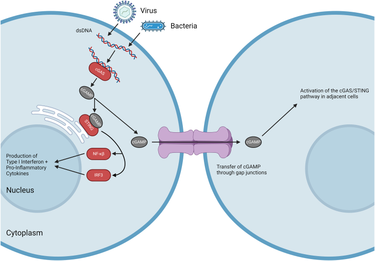
Connexins are essential for intercellular communication through gap junctions and the maintenance of cellular and tissue homeostasis. Connexin 43 (Cx43) is the most ubiquitously expressed connexin. As well as regulating homeostasis, Cx43 hemichannels and gap junctions play important roles in inflammation and the immune response. This, coupled with a range of non-channel functions performed by Cx43 makes it an attractive target for viruses. Recently, several groups have begun to explore the relationship between Cx43 and viral infection, with a diverse array of viruses being found to alter Cx43 hemichannels/gap junctions. Importantly, this includes several small DNA tumour viruses, which may target Cx43 to promote tumorigenesis. This review focuses on the ability of selected RNA/DNA viruses and retroviruses to either positively or negatively regulate Cx43 hemichannels and gap junctions in order to carry out their lifecycles. The role of Cx43 regulation by tumour viruses is also discussed in relation to tumour progression.

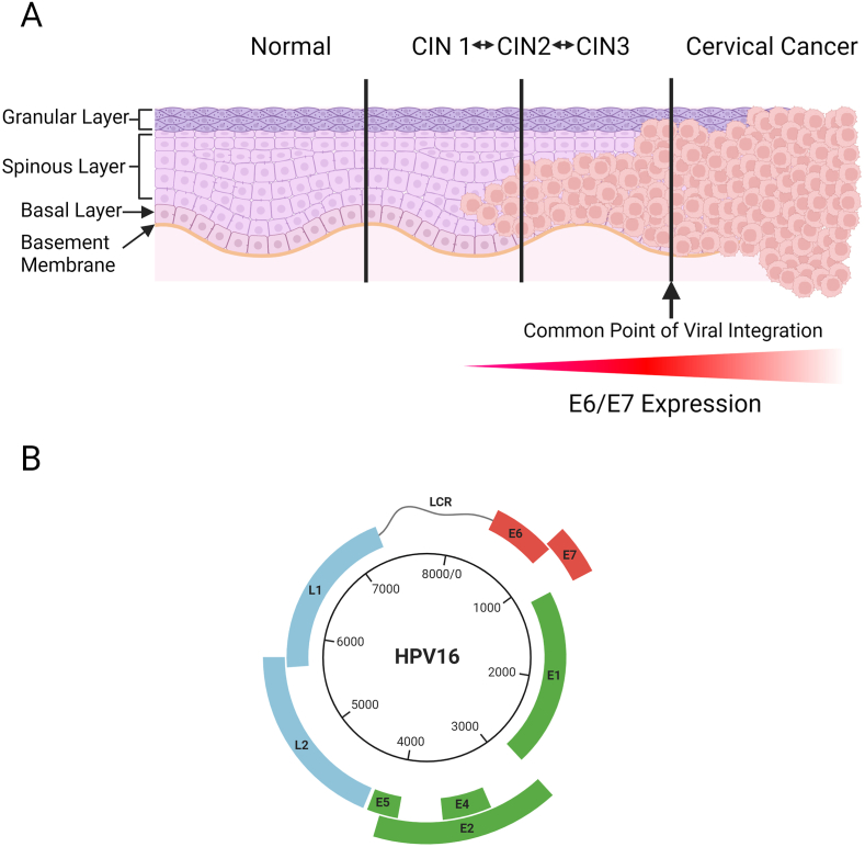
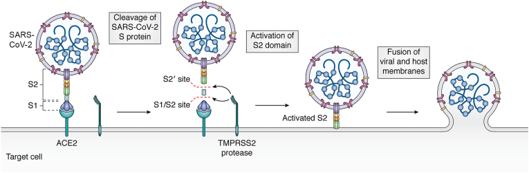
Keywords: Connexin 43; Gap junctions; Hemichannels; Human adenovirus type 5; Human immunodeficiency virus; Human papillomavirus; Severe acute respiratory syndrome coronavirus 2.


Conflict of interest statement

Declaration of competing interest The authors declare that they have no known competing financial interests or personal relationships that could have appeared to influence the work reported in this paper.

Figures

Fig. 1
Fig. 1
Structure of an individual Cx43 protein, a hemichannel (connexon) and a gap junction channel, with factors involved in inflammation and the immune response shown. Cx43 contains four α-helical transmembrane domains, two extracellular loops, a cytoplasmic loop and cytoplasmic N- and C-termini. A connexon is formed of six connexin proteins, which can be either homomeric or heteromeric depending on individual connexin protein compatibility [4]. Individual connexons inserted into the plasma membrane can act as hemichannels to release certain factors into the extracellular environment, including molecules involved in inflammation such as HMGB1, ATP and NAD+. Docking of two opposing connexons on the surfaces of different cells causes formation of a fully functional gap junction channel. These channels can also share factors involved in the immune response such as cGAMP for activation of the cGAS/STING pathway in adjacent cells and immunogenic peptides which can be presented on the surface of cells for recognition and killing of infected cells/cells adjacent to infected cells. Created in BioRender. Scott, H. (2024) BioRender.com/x05r178.
Fig. 2
Fig. 2
The role of gap junctions in the cGAS/STING Pathway. Sources of cytoplasmic dsDNA include bacteria, genomes of dsDNA viruses and DNA produced by reverse transcription of retroviral genomes. Other sources of dsDNA include dying cells and mitochondrial DNA (not shown). Binding of cytoplasmic dsDNA to cGAS leads to the generation of cyclic GMP-AMP (cGAMP) which is recognised by stimulator of interferon genes (STING). STING is shown on the endoplasmic reticulum membrane but also exists on the cell membrane and in the nucleus of cells. Through gap junctions, cGAMP is able to be passed to adjacent cells, allowing activation of the cGAS/STING pathway in cells without cytoplasmic dsDNA. Once bound by cGAMP, STING activates factors such as NF-кB and IRF3, which translocate to the nucleus to stimulate the production of pro-inflammatory cytokines and type I interferons. These responses lead to the production of factors which restrict pathogens through various mechanisms such as by inhibiting viral replication. Created in BioRender. Scott, H. (2024) BioRender.com/n18f996.
Fig. 3
Fig. 3
The HPV Genome and Cervical Cancer Progression. (A) Stages of cervical intraepithelial neoplasia (CIN) leading to cervical cancer following HPV infection. HPV infects the basal cells of the cervical epithelium through microabrasions. In these replicating basal cells, viral gene expression is low, with the productive viral lifecycle only taking place in the upper layers of the epithelium. In cells where the productive lifecycle takes place, HPV maintains cellular proliferation while inhibiting differentiation through expression of viral proteins. Infection with HPV can lead to the development of cervical intraepithelial neoplasia (CIN), which can be classified as CIN1, CIN2 or CIN3 depending on the proportion of the epithelium affected by HPV-related cell changes. The majority of CIN cases are cleared by the immune system within 24 months, however, if left untreated, a small proportion of these cases will progress to cervical cancer, which occurs when the undifferentiated persistently infected cells invade the basement membrane of the epithelium. Development of cervical cancer is heavily associated with integration of the viral genome into the host cell DNA and overexpression of the E6 and E7 oncoproteins. (B) The HPV16 genome is around 8 kB and encodes seven early regulatory proteins (E1, E2, E4, E5, E6, E7 and E8) and two late viral capsid proteins (L1 and L2), as well as possessing a long control region (LCR) which regulates viral transcription and replication. Upon infection of cells, the circular HPV genome is transported to the nucleus where it attaches to the host cell chromosomal DNA during cell division and is maintained in a circular (episomal) form. During cell division, the HPV genome is replicated with host cell DNA, being present in both resulting daughter cells. The HPV genome may linearise and integrate into the host cell genome following double-stranded DNA breaks, with this process resulting in a loss of the ability to produce infectious viral progeny and silencing of one or more viral genes. In the case of cancer, viral integration is also associated with unregulated expression of E6 and E7, which drive tumorigenesis. Created in BioRender. Scott, H. (2024) BioRender.com/n15a217.
Fig. 4
Fig. 4
Entry Route of SARS-CoV-2 into susceptible cells. The Spike protein of SARS-CoV-2 is composed of the S1 and S2 subunits, with S1 binding to the angiotensin converting enzyme 2 (ACE2) receptor, while the S2 subunit mediates fusion with the host membrane. On binding of S1 to ACE2, the TMPRSS2 cellular protease cleaves the spike protein at both the S1/S2 site and the S2′ site. This activates fusion activity of the S2 domain, which then allows fusion of the membranes, resulting in internalisation of the viral genome and the ACE2 receptor. Figure from Hartenian et al., 2020 [120].

Similar articles

Cited by

References

    1. Song B., Sheng X., Justice J.L., Lum K.K., Metzger P.J., Cook K.C., Kostas J.C., Cristea I.M. Intercellular communication within the virus microenvironment affects the susceptibility of cells to secondary viral infections. Sci. Adv. 2023;9 doi: 10.1126/SCIADV.ADG3433. - DOI - PMC - PubMed
    1. Yewdell J.W., Hill A.B. Viral interference with antigen presentation. Nat. Immunol. 2002;3(11):1019–1025. doi: 10.1038/ni1102-1019. 3 (2002) - DOI - PubMed
    1. Aasen T., Leithe E., Graham S.V., Kameritsch P., Mayán M.D., Mesnil M., Pogoda K., Tabernero A. Connexins in cancer: bridging the gap to the clinic. Oncogene. 2019;38:4429. doi: 10.1038/S41388-019-0741-6. - DOI - PMC - PubMed
    1. Koval M. Pathways and control of connexin oligomerization. Trends Cell Biol. 2006;16:159. doi: 10.1016/J.TCB.2006.01.006. - DOI - PMC - PubMed
    1. Wang N., De Bock M., Decrock E., Bol M., Gadicherla A., Vinken M., Rogiers V., Bukauskas F.F., Bultynck G., Leybaert L. Paracrine signaling through plasma membrane hemichannels. Biochim. Biophys. Acta. 2013;1828:35. doi: 10.1016/J.BBAMEM.2012.07.002. - DOI - PMC - PubMed

Publication types