. 2024 Nov 11;9(1):306.
doi: 10.1038/s41392-024-02011-y.

Overexpression of STX11 alleviates pulmonary fibrosis by inhibiting fibroblast activation via the PI3K/AKT/mTOR pathway

Affiliations

Overexpression of STX11 alleviates pulmonary fibrosis by inhibiting fibroblast activation via the PI3K/AKT/mTOR pathway

Guichuan Huang et al. Signal Transduct Target Ther. .

Abstract

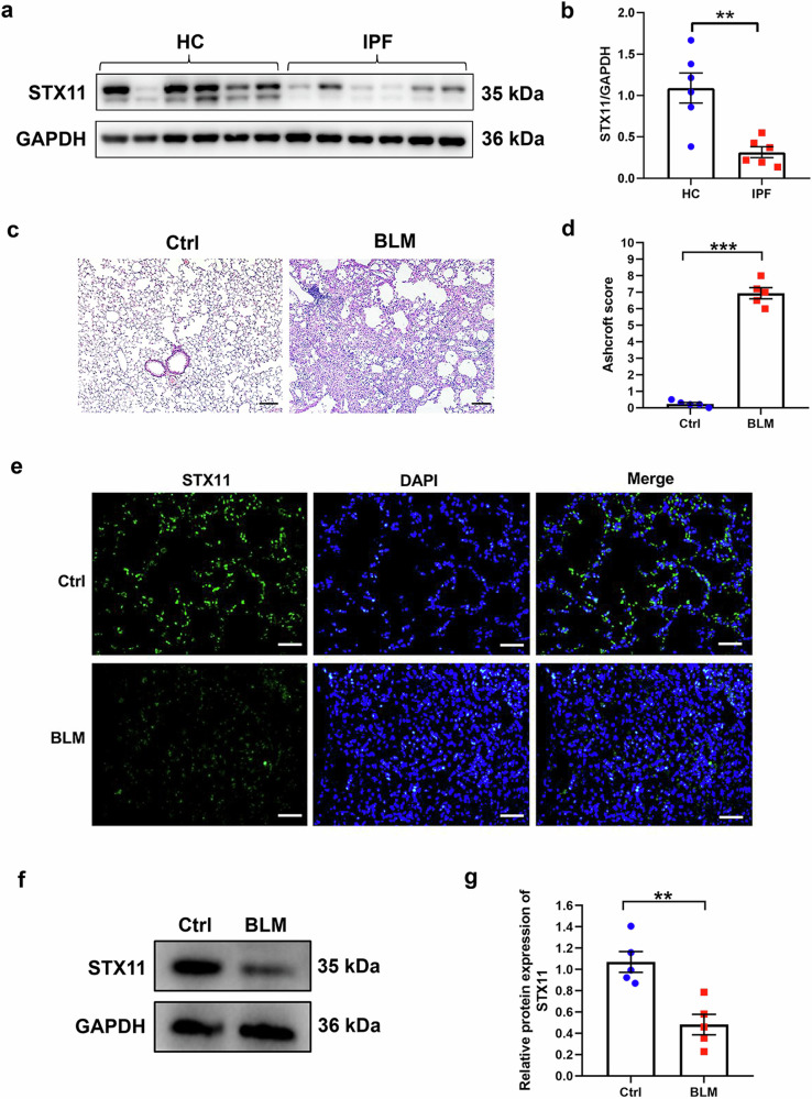
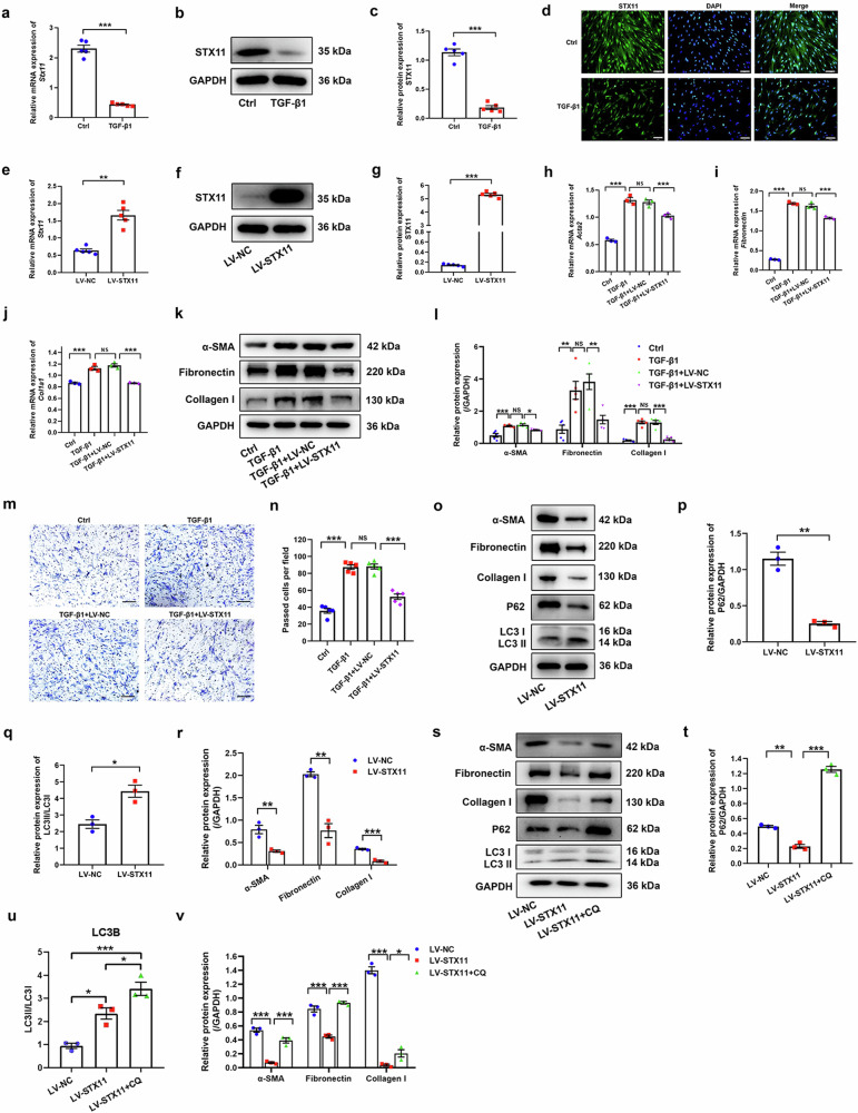
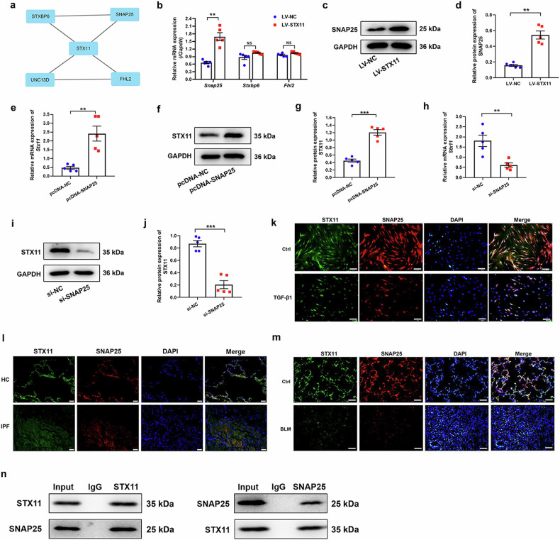
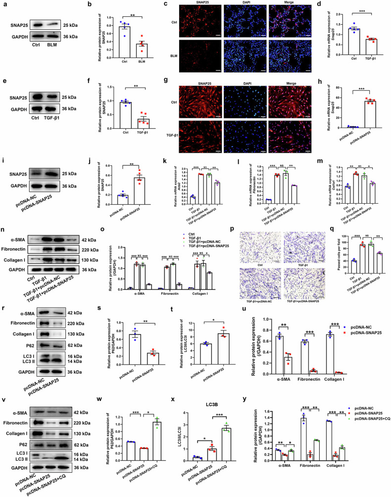
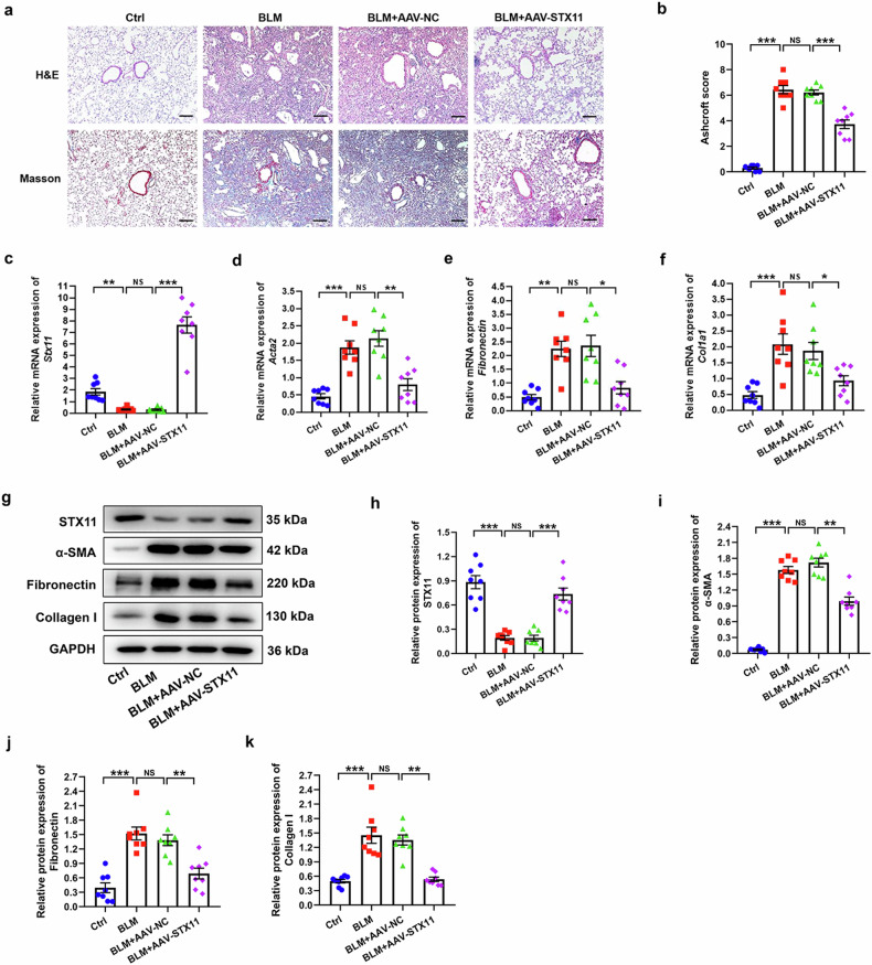
Fibroblast activation plays an important role in the occurrence and development of idiopathic pulmonary fibrosis (IPF), which is a progressive, incurable, and fibrotic lung disease. However, the underlying mechanism of fibroblast activation in IPF remains elusive. Here, we showed that the expression levels of STX11 and SNAP25 were downregulated in the lung tissues from patients with IPF and mice with bleomycin (BLM)-induced lung fibrosis as well as in the activated fibroblasts. Upregulation of STX11 or SNAP25 suppressed TGF-β1-induced activation of human lung fibroblasts (HLFs) via promoting autophagy. However, they failed to suppress fibroblast actviation when autophagy was blocked with the use of chloroquine (CQ). In addition, STX11 or SNAP25 could inhibit TGF-β1-induced fibroblast proliferation and migration. In vivo, overexpression of STX11 exerted its protective role in the mice with BLM-induced lung fibrosis. STX11 and SNAP25 mutually promoted expression of each other. Co-IP assay indicated that STX11 has an interaction with SNAP25. Mechanistically, STX11-SNAP25 interaction activated fibroblast autophagy and further inhibited fibroblast activation via blocking the PI3K/AKT/mTOR pathway. Overall, the results suggested that STX11-SNAP25 interaction significantly inhibited lung fibrosis by promoting fibroblast autophagy and suppressing fibroblast activation via blocking the PI3K/ATK/mTOR signaling pathway. Therefore, STX11 serves as a promising therapeutic target in IPF.


Conflict of interest statement

Competing interests The authors declare no competing interests.

Figures

Fig. 1
Fig. 1
The expression of STX11 in IPF and control lung tissues from the clinical specimen and mice. a, b The protein expression of STX11 in human IPF tissues and in normal human lung tissues was detected by western blot assay. c H&E staining was conducted on mice lung tissue sections from BLM group and control group. d The Ashcroft score was used to evaluate the degree of fibrosis in mice lung tissues. Immunofluorescence (e) and western blot (f, g) assays were applied to detect the expression of STX11 in mice lung tissues (original magnification ×200, scale bar: 100 μm). Data were expressed as mean ± SEM (n = 5 or 6). *p < 0.05; **p < 0.01; ***p < 0.001
Fig. 2
Fig. 2
The role of STX11 in fibroblast autophagy and activation. HLFs were treated with 10 ng/ml TGF-β1 for 48 h. a qPCR assay was used to detect the mRNA expression of STX11. b, c Western blot assay was used to detect the protein expression of STX11. d Immunofluorescence assay was used to detect the protein expression of STX11 (original magnification ×200, scale bar: 100 μm). HLFs were infected with lentiviruses harboring STX11 and treated with TGF-β1 for 48 h. qPCR (e) and western blot (f, g) assays were used to detect the expression of STX11. HLFs with STX11 overexpression were treated with 10 ng/ml TGF-β1 for 48 h. The expression of α-SMA, fibronectin, and COL1A1 (collagen I) was examined by qPCR (hj) and western blot (k, l) assays. m, n Transwell assay was used to determine the migration ability of HLFs (original magnification ×100, scale bar: 200 μm). or Western blot assay was used to determine the expression of fibroblast activation markers (α-SMA, fibronectin, collagen I) and autophagy-associated markers (LC3II/I, P62) in HLFs with STX11 overexpression. sv HLFs with STX11 overexpression were stimulated with chloroquine (CQ) at a final concentration of 10 mM for 48 h. Western blot assay was used to examine the expression of fibroblast activation markers and autophagy-associated markers in HLFs. GAPDH was used as an internal control. Data were expressed as mean ± SEM (n = 3 or 5). *p < 0.05; **p < 0.01; ***p < 0.001; NS, no significance
Fig. 3
Fig. 3
The relationship between STX11 and SNAP25. a The STRING online tool was used to identify the interaction protein with STX11. qPCR (b) and western blot (c, d) assays were used to detect the expression of SNAP25. qPCR (e) and western blot (f, g) assays were utilized to examine the expression of STX11 in HLFs transfected with pcDNA-NC or pcDNA-SNAP25. qPCR (h) and western blot (i, j) assays were conducted to explore the expression of STX11 in HLFs transfected with si-NC or si-SNAP25. k Immunofluorescence assay was performed to detect the expression of STX11 and SNAP25 in HLFs (original magnification ×200, scale bar: 100 μm). l Representative images showing STX11 (green), SNAP25 (red) and DAPI (blue) in HC and IPF lung sections by immunofluorescence staining (scale bar = 100 μm). m Immunofluorescence assay was performed to detect the expression of STX11 and SNAP25 in mice lung tissues (original magnification ×200, scale bar: 100 μm). n Reciprocal immunoprecipitation of STX11 and SNAP25 in HLFs. GAPDH was used as an internal control. Data were expressed as mean ± SEM (n = 5). **p < 0.01; ***p < 0.001
Fig. 4
Fig. 4
The role of SNAP25 in fibroblast autophagy and activation. a, b Western blot assay was used to detect the protein expression of SNAP25 in the mice lung. c Immunofluorescence assay was used to detect the protein expression of SNAP25 in the mice lung (original magnification ×200, scale bar: 100 μm). HLFs were treated with 10 ng/ml TGF-β1 for 48 h. qPCR (d) and western blot (e, f) assays were used to detect the expression of SNAP25 in HLFs. g Immunofluorescence assay was used to detect the protein expression of SNAP25 (original magnification ×200, scale bar: 100 μm). HLFs were transfected with pcDNA-NC or pcDNA-SNAP25 for 48 h. qPCR (h) and western blot (i, j) assays were conducted to identify the expression of SNAP25. HLFs were transfected with pcDNA-NC or pcDNA-SNAP25 for 24 h, and then stimulated with TGF-β1 for 48 h. qPCR (km) and western blot (n, o) assays were performed to determine the expression of α-SMA, fibronectin, and collagen I. p, q Transwell assay was used to determine the migration ability of HLFs (original magnification ×100, scale bar: 200 μm). ru Western blot assay was used to determine the expression of fibroblast activation markers (α-SMA, fibronectin, collagen I) and autophagy-associated markers (LC3II/I, P62) in HLFs with enhanced expression of SNAP25. vy HLFs with SNAP25 overexpression were stimulated with chloroquine (CQ) at a final concentration of 10 mM for 48 h. Western blot assay was used to detect the expression of fibroblast activation markers and autophagy-associated markers in HLFs. GAPDH was used as an internal control. Data were expressed as mean ± SEM (n = 3 or 5). *p < 0.05; **p < 0.01 ; ***p < 0.001
Fig. 5
Fig. 5
The effect of STX11 and SNAP25 on the PI3K/AKT/mTOR pathway. a, b HLFs with STX11 overexpression were transfected with si-NC or si-SNAP25 for 24 h, and then stimulated with TGF-β1 for 48 h. Western blot assay was performed to determine the expression of α-SMA, fibronectin, and collagen I. cf The expression of the PI3K/AKT/mTOR pathway related proteins was detected by western blot in HLFs with STX11 overexpression. gj HLFs with forced expression of STX11 were pretreated with 100 ng/ml IGF-1 for 1 h, and exposed to TGF-β1 for 48 h. Western blot assay was utilized to detect the protein expression in HLFs. kn The expression of the PI3K/AKT/mTOR pathway related proteins was detected by western blot in HLFs with SNAP25 overexpression. or HLFs with forced expression of SNAP25 were pretreated with 100 ng/ml IGF-1 for 1 h, and exposed to TGF-β1 for 48 h. Western blot assay was utilized to detect the protein expression in HLFs. sv HLFs with SNAP25 silencing were pretreated with 10 μM LY294002 for 1 h, and exposed to TGF-β1 for 48 h. Western blot assay was utilized to detect the protein expression in HLFs. Data were expressed as mean ± SEM (n = 3 or 5). *p < 0.05; **p < 0.01 ; ***p < 0.001; NS, no significance
Fig. 6
Fig. 6
Overexpression of STX11 inhibits BLM-induced pulmonary fibrosis in mice. a Representative images of H&E and Masson staining in the lung sections of mice. b Ashcroft score based on H&E staining was determined. cf The mRNA expression of STX11, α-SMA, fibronectin, and COL1A1 was detected by qPCR. gk The protein expression of STX11, α-SMA, fibronectin, and collagen I was detected by western blot. GAPDH was used as an internal control. Data were expressed as mean ± SEM (n = 8). *p < 0.05; **p < 0.01; *** p < 0.001; NS, no significance
Fig. 7
Fig. 7
Schematic diagram of the study. STX11 forms a complex with SNAP25, blocking the PI3K/AKT/mTOR pathway and promoting autophagy of lung fibroblasts, which inhibits the development of lung fibrosis

References

    1. Barratt, S. L., Creamer, A., Hayton, C. & Chaudhuri, N. Idiopathic Pulmonary Fibrosis (IPF): An Overview. J. Clin. Med.7, 201 (2018). - PMC - PubMed
    1. Yu, Q. Y. & Tang, X. X. Irreversibility of pulmonary fibrosis. Aging Dis.13, 73–86 (2022). - PMC - PubMed
    1. King, T. E. et al. All-cause mortality rate in patients with idiopathic pulmonary fibrosis. Implications for the design and execution of clinical trials. Am. J. Respir. Crit. Care Med.189, 825–831 (2014). - PubMed
    1. Hong, W. & Lev, S. Tethering the assembly of SNARE complexes. Trends Cell Biol.24, 35–43 (2014). - PubMed
    1. Tang, B. L. SNAREs and developmental disorders. J. Cell Physiol.236, 2482–2504 (2021). - PubMed

Publication types

MeSH terms

LinkOut - more resources