. 2024 Nov 10;14(1):27445.
doi: 10.1038/s41598-024-78380-9.

Vitamin D receptor and its antiproliferative effect in human pulmonary arterial hypertension

Affiliations

Vitamin D receptor and its antiproliferative effect in human pulmonary arterial hypertension

Maria Callejo et al. Sci Rep. .

Abstract

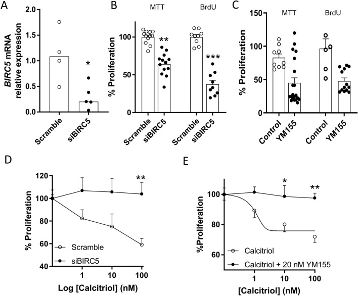
Vitamin D (vitD) deficiency is frequently observed in patients with pulmonary arterial hypertension (PAH) and, in these patients, low levels of vitD correlate with worse prognosis. The aim of this study was to examine the expression and the antiproliferative role of vitD receptor (VDR) and its signalling pathway in the human pulmonary vasculature. VDR presence and expression was analyzed in lungs, pulmonary artery smooth muscle cells (PASMC) and endothelial cells (PAEC) from controls and PAH-patients. VDR expression and VDR-target genes were examined in PASMC treated with calcitriol. The antiproliferative effect of 48 h-calcitriol was studied in PASMC by MTT and BrdU assays. VDR is expressed in PASMC. It is downregulated in lungs and in PASMC, but not in PAEC, from PAH-patients compared to non-hypertensive controls. Calcitriol strongly upregulated VDR expression in PASMC and the VDR target genes KCNK3 (encoding TASK1), BIRC5 (encoding survivin) and BMP4. Calcitriol produced an antiproliferative effect which was diminished by silencing or by pharmacological inhibition of survivin or BMPR2, but not of TASK1. In conclusion, the expression of VDR is low in PAH-patients and can be rescued by calcitriol. VDR exerts an antiproliferative effect in PASMC by modulating survivin and the BMP signalling pathway.


Conflict of interest statement

Declarations Competing interests The authors declare no competing interests. Ethical approval Research using human samples were approved by the Ethics Committee of the Getafe Hospital (Madrid, Spain), Ciberes Biobank (Barcelona, Spain) and French institutional Ethics Committee (Protocol N8CO-08-003, ID RCB: 2008-A00485-50). Informed consent was obtained in all cases. Human explanted lung tissues from non-PAH subjects were obtained from non-tumor lung areas of the resection specimens from patients undergoing surgery for lung carcinoma or discarded for lung transplantation. The donor lungs included in this study were thoroughly reviewed by a pathologist. PAH samples were obtained from lung transplantation.

Figures

Fig. 1
Fig. 1
VDR is relatively highly expressed in human lungs and it is downregulated in PAH. (A) VDR gene expression pattern in different tissues. Data from the NCBI database, project PRJEB4337. Data is log-transformed into Log2(RPKM + 1) and represented as dot plot and mean. (B) Analysis of VDR mRNA expression in transcriptome database GSE117261 in controls and PAH-patients (All PAH). The analysis was also segregated by sex and by subclass of PAH, including associated (APAH), familial PAH (FPAH), idiopathic PAH (IPAH) or other or unknown causes. (C) VDR mRNA expression by qRT-PCR and (D) VDR protein normalized by GAPDH expression by Western Blot in lungs samples from controls and PAH-patients. Multiple bands of VDR were quantified according to the manufacturer’s antibody technical sheet. Results are represented as scatter plots and bars with medians. **p < 0.01 and ***p < 0.001, non-parametric Mann Whitney test.
Fig. 2
Fig. 2
VDR is expressed in cultured human PASMC and it is upregulated by calcitriol. (A) Representative immunohistochemistry of VDR in a control human lung showing a pulmonary artery, the elastin autofluorescence is shown in green. (B) Uniform manifold approximation and projection (UMAP) plot of the pulmonary artery scRNA-Seq data of donor (Control, n = 3) and pulmonary arterial hypertension (PAH, n = 3) samples are colored by cell identity. (C) Dot plot with VDR gene expression by cell identity and condition (Control and PAH). (D) Immunocytochemistry of PASMC stained with anti-VDR. (EG) PASMC VDR mRNA expression by qRT-PCR in (E) PAEC (n = 9) vs PASMC (n = 6) from control donors, (F) PASMC from control donors (n = 6) vs PASMC from PAH patients (n = 8), and (G) PAEC from control donors (n = 9) vs PAEC from PAH patients (n = 13). (H) VDR upregulation at 24 and 48 h by calcitriol (100 nmol/l) in PASMC from control donors (n = 6) and PASMC from PAH patients (n = 8). Results are represented as scatter plots and bars with medians. ***p < 0.001 vs PAEC, *p < 0.05 vs controls, non-parametric Mann Whitney test. In panel H, the box shows two-way ANOVA results and **p < 0.01 following Bonferroni’s multiple comparisons test.
Fig. 3
Fig. 3
Calcitriol exerts antiproliferative effects in human PASMC. (A) and (B) Proliferation in human PASMC from 5 (in triplicate) controls measured by BrdU incorporation and MTT assay, respectively, and (C) proliferation in PASMC from 4 different cultures from PAH-patients, in duplicate or triplicate, by MTT assay, after exposure to calcitriol (1–100 nmol/l) for 48 h. Results are expressed as mean ± SEM. *, **, *** indicates p < 0.05, p < 0.01 and p < 0.001 vs vehicle, one-way ANOVA, Bonferroni test.
Fig. 4
Fig. 4
Calcitriol modulates the expression of genes of interest in PAH. Expression of (A) KCNK3 (gene encoding TASK1, n = 6), (B) BIRC5 (gene encoding survivin, n = 7) and (C) Genes involved in the BMP signaling pathway (n = 3–6) after exposure to vehicle o calcitriol (100 nmol/l) for 48 h. Data in panels A and B are represented as plot before-after. The grey point represents the mean. *p < 0.05, non-parametric paired test vs vehicle. Results in panel C are expressed as mean ± SEM.
Fig. 5
Fig. 5
Calcitriol inhibits PASMC proliferation via BMPR2. Human control PASMC were transfected with siRNA for BMPR2 (siBMPR2) or control siRNA (scramble). (A) BMPR2 mRNA expression assessed by qRT-PCR after 48 h post-transfection. Data is expressed as scatter plots and bars with medians, *p < 0.05, Mann–Whitney test. (B) Effects of calcitriol on proliferation in silenced BMPR2 PASMC measured by MTT assay. Results are expressed as mean ± SEM. *p < 0.05, t-test vs scramble. (C) and (D) Effects of calcitriol (1–100 nmol/l) on proliferation in PASMC transfected with siBMPR2 or scramble, measured by MTT and BrdU assays, respectively. *p < 0.05, two-way ANOVA, Bonferroni post hoc test, vs scramble. (E) Effects of calcitriol on proliferation in PASMC in the presence or absence of DMH1 (5 µmol/l), by MTT and BrdU assay; ***p < 0.001 calcitriol vs vehicle (black column), two way-ANOVA. n = 4–5 different cultures in duplicate or triplicate.
Fig. 6
Fig. 6
The antiproliferative effect of calcitriol is suppressed in the presence of survivin inhibition. (A) BIRC5 (gene encoding survivin) mRNA expression assessed by qRT-PCR 48 h post-transfection with siRNA against BIRC5 (siBIRC5) or scramble siRNA. Data is expressed as scatter plots and bars represent the median, *p < 0.05, Mann–Whitney test. (B, C) Proliferation of PASMC transfected with siBIRC5 or scramble (B) or YM155 (20 nmol/l) measured by MTT and BrdU assays. (D, E) Effects of calcitriol (1–100 nmol/l) on proliferation in PASMC transfected with siBIRC5 or scramble (D) or treated with YM155 (20 nmol/l) (E) measured by BrdU assay for 48 h. Data are expressed as mean ± SEM. *p < 0.05 and **p < 0.01 vs scramble or calcitriol, two-way ANOVA, Bonferroni test. n = 3–4 different cultures in triplicate.

Similar articles

Cited by

References

    1. Simonneau, G. et al. Haemodynamic definitions and updated clinical classification of pulmonary hypertension. Eur. Respir. J.53, 1801913. 10.1183/13993003.01913-2018 (2019). - PMC - PubMed
    1. Wilkins, M. R. Pulmonary hypertension: The science behind the disease spectrum. Eur. Respir. Rev.21, 19–26. 10.1183/09059180.00008411 (2012). - PMC - PubMed
    1. Pullamsetti, S. S., Savai, R., Seeger, W. & Goncharova, E. A. Translational advances in the field of pulmonary hypertension. From cancer biology to new pulmonary arterial hypertension therapeutics. Targeting cell growth and proliferation signaling hubs. Am. J. Respir. Crit. Care Med.195, 425–437. 10.1164/rccm.201606-1226PP (2017). - PMC - PubMed
    1. Callejo, M. et al. Total, bioavailable, and free vitamin d levels and their prognostic value in pulmonary arterial hypertension. J. Clin. Med.9, 448. 10.3390/jcm9020448 (2020). - PMC - PubMed
    1. Tanaka, H. et al. Therapeutic impact of dietary vitamin D supplementation for preventing right ventricular remodeling and improving survival in pulmonary hypertension. PLoS One12, e0180615. 10.1371/journal.pone.0180615 (2017). - PMC - PubMed

MeSH terms

LinkOut - more resources