. 2025 Jan 1;29(1):93-108.
doi: 10.4196/kjpp.24.265. Epub 2024 Nov 11.

The mutual interaction of TRPC5 channel with polycystin proteins

Affiliations

The mutual interaction of TRPC5 channel with polycystin proteins

Misun Kwak et al. Korean J Physiol Pharmacol. .

Abstract

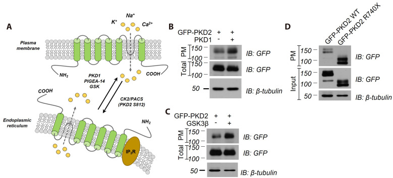
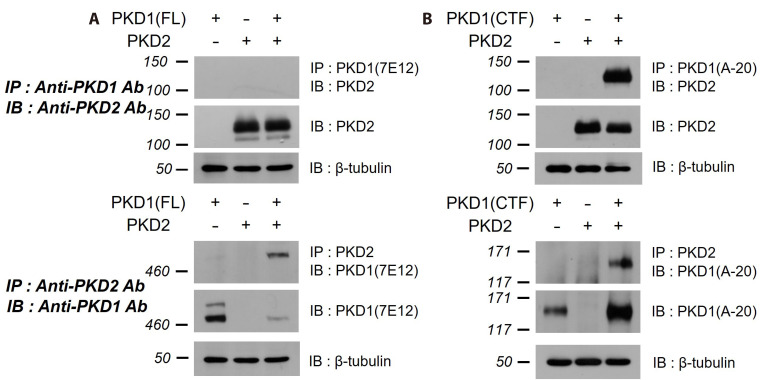
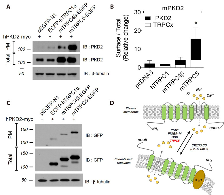
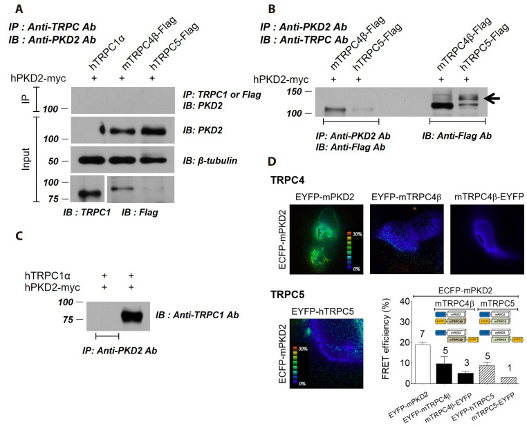
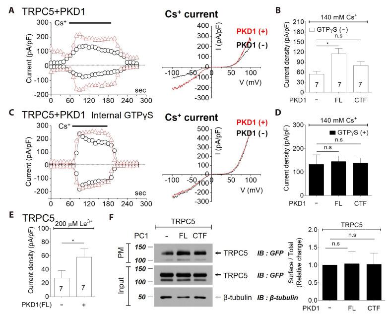
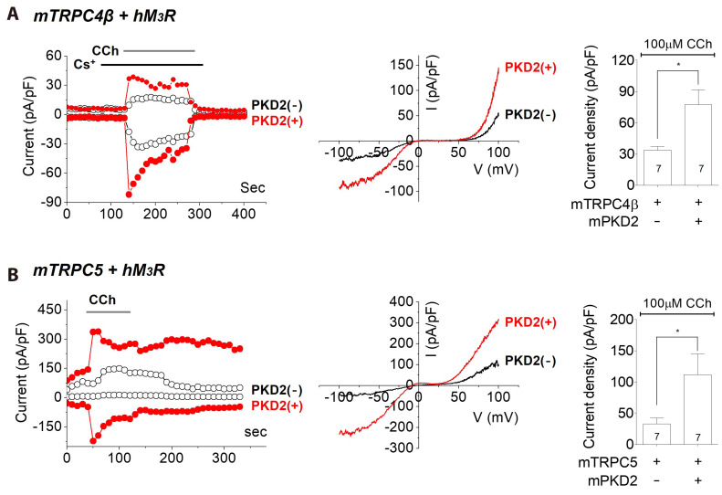
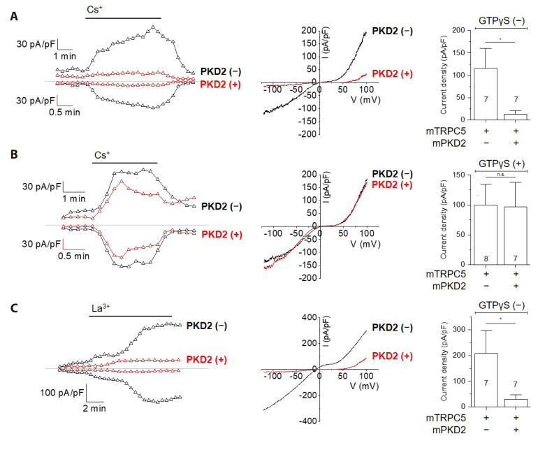
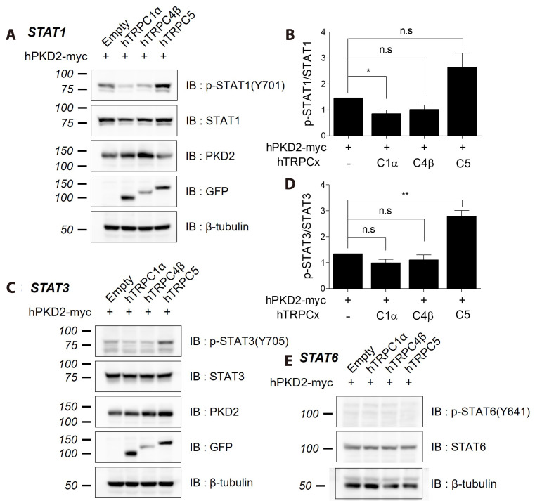
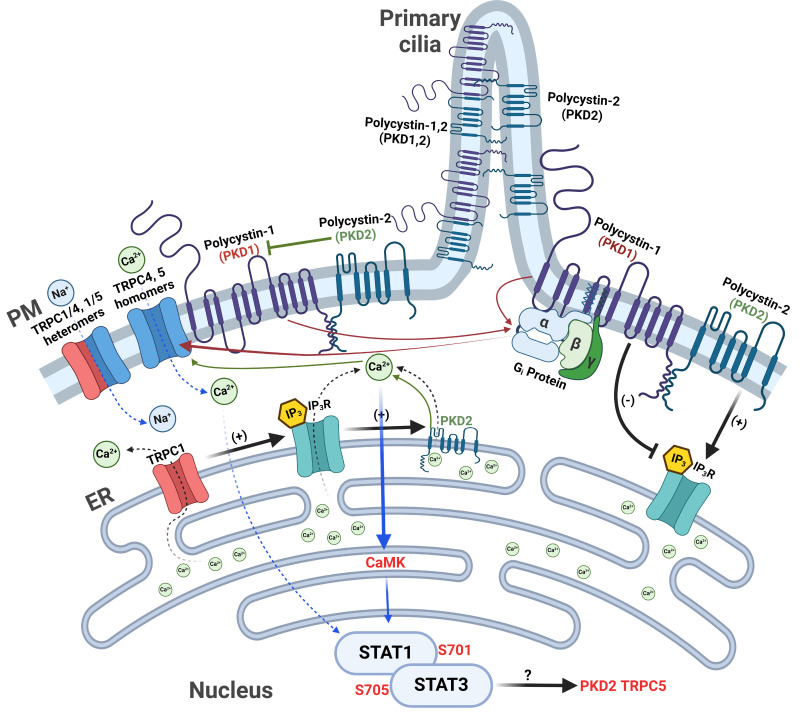
PKD1 regulates a number of cellular processes through the formation of complexes with the PKD2 ion channel or transient receptor potential classical (TRPC) 4 in the endothelial cells. Although Ca2+ modulation by polycystins has been reported between PKD1 and TRPC4 channel or TRPC1 and PKD2, the function with TRPC subfamily regulated by PKD2 has remained elusive. We confirmed TRPC4 or TRPC5 channel activation via PKD1 by modulating G-protein signaling without change in TRPC4/C5 translocation. The activation of TRPC4/C5 channels by intracellular 0.2 mM GTPγS was not significantly different regardless of the presence or absence of PKD1. Furthermore, the C-terminal fragment (CTF) of PKD1 did not affect TRPC4/C5 activity, likely due to the loss of the N-terminus that contains the G-protein coupled receptor proteolytic site (GPS). We also investigated whether TRPC1/C4/C5 can form a heterodimeric channel with PKD2, despite PKD2 being primarily retained in the endoplasmic reticulum (ER). Our findings show that PKD2 is targeted to the plasma membrane, particularly by TRPC5, but not by TRPC1. However, PKD2 did not coimmunoprecipitate with TRPC5 as well as with TRPC1. PKD2 decreased both basal and La3+-induced TRPC5 currents but increased M3R-mediated TRPC5 currents. Interestingly, PKD2 increased STAT3 phosphorylation with TRPC5 and decreased STAT1 phosphorylation with TRPC1. To be specific, PKD2 and TRPC1 compete to bind with TRPC5 to modulate intracellular Ca2+ signaling and reach the plasma membrane. This interaction suggests a new therapeutic target in TRPC5 channels for improving vascular endothelial function in polycystic kidney disease.

Keywords: Polycystic kidney disease 1 protein; Polycystic kidney disease 2 protein; Polycystic kidney diseases; Signal transducer and activator of transcription; Transient receptor potential canonical channel 5.


Conflict of interest statement

CONFLICTS OF INTEREST

The authors declare no conflicts of interest.

Figures

Fig. 1
Fig. 1. The effects of PKD1 and GSK3
β on PKD2 expression. (A) Schematic illustration of factors affecting the trafficking of PKD2 between the endoplasmic reticulum (ER) and the plasma membrane (PM). (B, C) Surface expression of PKD2-GFP in HEK cells coexpressing PKD1 (B) or GSK3β (C). The surface expression of PKD2 was increased by PKD1 or GSK3β, as determined by co-expression and surface biotinylation. (D) Deletion of the PKD2 ER retention signal (PKD2 R740X) increased the surface expression of PKD2. Surface and total PKD2 were detected by immunoblotting with an anti-GFP antibody. PKD, polycystic kidney disease; IB, immunoblot.
Fig. 2
Fig. 2. The interaction of hPKD1 CTF with hPKD2-myc.
PKD1(FL or CTF) flag-tagged at the C-terminus was coexpressed with PKD2. PKD1/PKD2 complex was immunoprecipitated using an antibody against the PKD1 N-terminus (7E12) (A upper panel) or an antibody against the PKD1 C-terminus (A-20) (B upper panel) and detected by anti-PKD2 antibody (H-280). PKD1/PKD2 complex was also immunoprecipitated using anti-PKD2 antibody (H-280) and detected an antibody against the PKD1 N-terminus (7E12) (A lower panel) or an antibody against the PKD1 C-terminus (A-20) (B lower panel). FL and NTF forms of PKD1 were observed with FL-Flag. The cleaved CTF forms and non-cleaved FL were detected by an anti-PKD1 (A-20) antibody. PKD1 FL or CTF co-immunoprecipitated with PKD2 but not PKD1 NTF. PKD, polycystic kidney disease; CTF, C-terminal fragment; FL, full-length; NTF, N-terminal fragment; IP, immunoprecipitation; IB, immunoblot.
Fig. 3
Fig. 3. The effects of TRPC on the surface expression of PKD2.
(A) The surface expression of PKD2 in HEK cells coexpressing TRPC. Surface expression of PKD2-myc was increased by TRPC5 as determined by co-expression and surface biotinylation. Immunoblots of surface and total were detected by anti-PKD2 antibody (H-280). (B) Bar graphs showing mean levels of surface PKD2 relative to total PKD2 protein levels (black bar, n = 5), and mean levels of surface TRPC relative to total TRPC protein levels (white bar, n = 5). Statistical significance is denoted by an asterisk (*p < 0.05). (C) The surface expression of TRPC in HEK cells coexpressing PKD2. Surface expression of TRPC5 was increased by PKD2 as determined by co-expression and surface biotinylation. Immunoblots of surface and total were detected by anti-GFP antibody. (D) The schematic illustration of factors affecting the trafficking of PKD2 between the endoplasmic reticulum and the plasma membrane (PM). TRPC5 increases the surface expression of PKD2. TRPC, classical transient receptor potential; PKD, polycystic kidney disease; IB, immunoblot.
Fig. 4
Fig. 4. The interactions between TRPC and PKD2.
(A) HEK cells were transfected with TRPC1α, TRPC4β-Flag, or TRPC5-Flag along with PKD2-Myc and were used to test co-immunoprecipitation (Co-IP) of TRPC and PKD2. Anti-TRPC1 or anti-Flag antibody was used for immunoprecipitation (IP), and anti-PKD2 antibody was used for detecting PKD2-Myc. TRPC did not Co-IP with PKD2. (B, C) Anti-PKD2 antibody (H-280) was used for IP. TRPC4 or TRPC5 was detected with anti-Flag antibody (B), and TRPC1 was detected with anti-TRPC1 antibody (C). TRPC and PKD2 were not co-immunoprecipitated reciprocally. (D) FRET between TRPC and PKD2. FRET-detectable interactions occur between EYFP-PKD2 and ECFP-PKD2. Representative FRET images of ECFP-PKD2 co-expressed with EYFP-PKD2, EYFP-TRPC4, and TRPC4-EYFP are shown in the upper panel. In the lower panel, FRET images of ECFP-PKD2 co-expressed with EYFP-TRPC5 and a bar graph of FRET efficiency between PKD2 and TRPC are shown. TRPC, classical transient receptor potential; PKD, polycystic kidney disease; FRET, fluorescence resonance energy transfer; IB, immunoblot.
Fig. 5
Fig. 5. The effects of PKD1 on TRPC5 channels.
(A) HEK 293 cells were co-transfected with TRPC5 and PKD(FL). The TRPC5 current was recorded without GTPγS, and the current amplitude was increased by 140 mmol/L Cs+. The current amplitudes at −60 mV and +60 mV were plotted against time (the left panel) in HEK cells expressing PKD1 and TRPC5 (red triangle) or TRPC5 only (black circle). The ramp pulses were applied every 10 sec. The I-V curves from HEK cells expressing PKD1 and TRPC5 (red line) or TRPC5 only (black line) showed a double-rectifying shape. (B) The bar graphs represent the means ± S.E.M of current density (pA/pF) at −60 mV in the absence of GTPγS infusion. (C) The current was recorded in TRPC5/PKD1 co-transfected HEK 293 cells infused with GTPγS. The I-V curves from HEK cells expressing PKD1 and TRPC5 (red line) or TRPC5 only (black line) showed a double-rectifying shape. (D) The bar graphs represent the means ± S.E.M of current density (pA/pF) at −60 mV in the presence of GTPγS infusion. (E) The effect of PKD1 on La3+-induced TRPC5 current. (F) The surface expression of TRPC5 in HEK cells coexpressing PKD1. Surface expression of TRPC5 was not increased by PKD1 as determined by co-expression and surface biotinylation. Immunoblots of surface and total were detected by anti-GFP antibody. Right panel: Bar graph showing the effect of PKD1 on the surface expression of TRPC5 (n = 3). *p < 0.05. PKD, polycystic kidney disease; TRPC, classical transient receptor potential, FL, full-length; CTF, C-terminal fragment; PM, plasma membrane; IB, immunoblot; n.s., not significant.
Fig. 6
Fig. 6. The effects of PKD2 on TRPC4 and TRPC5 channels stimulated by carbachol.
(A) HEK 293 cells were co-transfected with TRPC4 and PKD2, and M3 receptor was also expressed. TRPC4 current was activated by carbachol. Current amplitudes at −60 mV and +60 mV were plotted against time (left panel) in HEK cells expressing PKD2 and TRPC4 (red circles) or TRPC4 only (black circles). Ramp pulses were applied every 10 sec. I-V curves from HEK cells expressing PKD2 and TRPC4 (red line) or TRPC4 only (black line) showed a double-rectifying shape (right panel). Bar graphs represent the mean ± S.E.M of current density (pA/pF) at −60 mV. (B) HEK 293 cells were co-transfected with TRPC5 and PKD2, and M3 receptor was also expressed. TRPC5 current was activated by carbachol. Current amplitudes at −60 mV and +60 mV were plotted against time (left panel) in HEK cells expressing PKD2 and TRPC5 (red circles) or TRPC5 only (black circles). Ramp pulses were applied every 10 sec. I-V curves from HEK cells expressing PKD2 and TRPC5 (red line) or TRPC5 only (black line) showed a double-rectifying shape (right panel). Bar graphs represent the mean ± S.E.M of current density (pA/pF) at −60 mV. PKD, polycystic kidney disease; TRPC, classical transient receptor potential; CCh, carbachol.
Fig. 7
Fig. 7. The effects of PKD2 on basal and La3+ induced TRPC5 currents.
(A) TRPC5 basal current without GTPγS. HEK 293 cells were co-transfected with TRPC5 and PKD2. The basal current of TRPC5 channels was increased by external cesium. The current amplitudes at −60 mV and +60 mV were plotted against time (the left panel) in HEK cells expressing PKD2 and TRPC5 (red triangle) or TRPC5 only (black triangle). The ramp pulses were applied every 10 sec. The I-V curves from HEK cells expressing PKD2 and TRPC5 (red line) or TRPC5 only (black line) showed a double-rectifying shape. (right panel) The bar graphs represent the means ± S.E.M of current density (pA/pF) at −60 mV. (B) TRPC5 activated with GTPγS. HEK 293 cells were co-transfected with TRPC5 and PKD2. The TRPC5 current was activated by GTPγS and external cesium. The current amplitudes at −60 mV and +60 mV were plotted against time (the left panel) in HEK cells expressing PKD2 and TRPC5 (red triangle) or TRPC5 only (black triangle). The ramp pulses were applied every 10 sec. The I-V curves from HEK cells expressing PKD2 and TRPC5 (red line) or TRPC5 only (black line) showed a double-rectifying shape. (right panel) The bar graphs represent the means ± S.E.M of current density (pA/pF) at −60 mV. (C) La3+ induced TRPC5 current. The TRPC5 current was activated by external La3+. The current amplitudes at −60 mV and +60 mV were plotted against time (the left panel) in HEK cells expressing PKD2 and TRPC5 (red triangle) or TRPC5 only (black triangle). The ramp pulses were applied every 10 sec. The I-V curves from HEK cells expressing PKD2 and TRPC5 (red line) or TRPC5 only (black line) showed a double-rectifying shape. (right panel) The bar graphs represent the means ± S.E.M of current density (pA/pF) at −60 mV. PKD, polycystic kidney disease; TRPC, classical transient receptor potential.
Fig. 8
Fig. 8. STAT phosphorylation by coexpression of PKD2 with TRPC.
(A) Effects of TRPC on STAT1 phosphorylation. HEK 293 cells were transfected with PKD2 and TRPC. Cell lysates were subjected to Western blot analysis using antibodies against total and phosphorylated STAT1. (B) Bar graphs showing mean levels of phosphorylated STAT1 relative to total STAT1 protein levels (n = 5). Statistical significance is denoted by an asterisk (*p < 0.05). The bar graphs represent the means ± S.E.M. (C) Effects of TRPC on STAT3 phosphorylation. HEK 293 cells were transfected with PKD2 and TRPC. Cell lysates were subjected to Western blot analysis using antibodies against total and phosphorylated STAT3. (D) Bar graphs showing mean levels of phosphorylated STAT3 relative to total STAT3 protein levels (n = 5). Statistical significance is denoted by an asterisk (**p < 0.01). (E) Effects of TRPC on STAT6 phosphorylation. STAT, signal transducer and activator of transcription; PKD, polycystic kidney disease; TRPC, classical transient receptor potential; n.s., not significant; IB, immunoblot.
Fig. 9
Fig. 9. Schematic illustration of TRPC, PKD, and IP3R interaction at the endoplasmic reticulum (ER) and their trafficking to the plasma membrane (PM).
The schematic illustration of the interaction between TRPC and PKD proteins via the ER. TRPC1 and PKD2 function as calcium release channels in the ER. IP3R interacts with both TRPC1 and PKD2 at the ER. The red arrow shows how PKD1 regulates TRPC function. The green arrow shows the process of regulating TRPC through PKD2. The blue arrow shows the signaling process through TRPC. PKD1 acts as a GPCR and activates Gαi proteins. The activated Gαi proteins induced TRPC4 and TRPC5 currents. On the other hand, PKD2 binds to PKD1 and inhibits the activity of PKD1, which lowers the basal activity of G protein and inhibits the basal current and La3+-induced current of TRPC5. Interestingly, when activated through M3R, calcium release through IP3R is added, and the function of PKD2 changes to activating TRPC5 current. This is because PKD2 acts as a calcium-releasing ion channel in the ER, and IP3R further promotes PKD2 function, and vice versa. When TRPC4/5 is activated, calcium is supplied abundantly into the cell, and STAT1 (S701 phosphorylation) and STAT3 (S705 phosphorylation) is activated through the increased calcium and CaMK. STAT phosphorylation might increase the surface expression of PKD2 and TRPC5. TRPC, classical transient receptor potential; PKD, polycystic kidney disease; GPCR, G protein coupled receptor; STAT, signal transducer and activator of transcription.

Similar articles

References

    1. Torres VE, Harris PC, Pirson Y. Autosomal dominant polycystic kidney disease. Lancet. 2007;369:1287–1301. doi: 10.1016/S0140-6736(07)60601-1. - DOI - PubMed
    1. Torres VE, Harris PC. Progress in the understanding of polycystic kidney disease. Nat Rev Nephrol. 2019;15:70–72. doi: 10.1038/s41581-018-0108-1. - DOI - PMC - PubMed
    1. Yamaguchi T, Hempson SJ, Reif GA, Hedge AM, Wallace DP. Calcium restores a normal proliferation phenotype in human polycystic kidney disease epithelial cells. J Am Soc Nephrol. 2006;17:178–187. doi: 10.1681/ASN.2005060645. - DOI - PubMed
    1. Cabrita I, Talbi K, Kunzelmann K, Schreiber R. Loss of PKD1 and PKD2 share common effects on intracellular Ca2+ signaling. Cell Calcium. 2021;97:102413. doi: 10.1016/j.ceca.2021.102413. - DOI - PubMed
    1. Nilius B, Owsianik G, Voets T, Peters JA. Transient receptor potential cation channels in disease. Physiol Rev. 2007;87:165–217. doi: 10.1152/physrev.00021.2006. - DOI - PubMed