

Coexistence of uterine adenosarcoma and endometrioid endometrial carcinoma: A case report and literature review
- PMID: 39523612
- PMCID: PMC11552035
- DOI: 10.1177/00368504241296291
Coexistence of uterine adenosarcoma and endometrioid endometrial carcinoma: A case report and literature review
Abstract
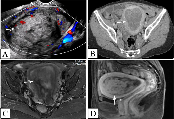
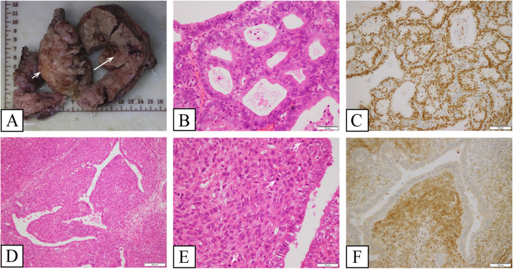
Uterine adenosarcoma coexisting with endometrial carcinoma is a very rare disease. Herein, we reported the case of uterine adenosarcoma coexisting with endometrioid endometrial carcinoma. Transvaginal ultrasound, computed tomography, and magnetic resonance imaging examinations all indicated a space-occupying lesion in the uterine cavity, and initially was considered endometrial carcinoma. Subsequently, total hysterectomy combined with bilateral salpingo-oophorectomy, pelvic lymphadenectomy, and para-aortic lymphadenectomy were performed. The coexistence of uterine adenosarcoma and endometrioid endometrial carcinoma was histologically confirmed postoperatively. The patient recovered well after surgery and was discharged on postoperative day 7. At a follow-up examination 10 months after surgery, we found no evidence of discomforting symptoms and recurrence or metastasis. Since the coexistence of uterine adenosarcoma and endometrial carcinoma is rare, it is easy to be overlooked the presence of uterine adenosarcoma on imaging or morphology, and thus be misdiagnosed as a more common disease, namely endometrial carcinoma. Observing the cystic structure within the lesion on magnetic resonance imaging is helpful for the diagnosis of uterine adenosarcoma. This article summarizes the imaging characteristics, clinicopathological features, molecular correlation, treatment, and prognosis of the disease.
Keywords: Uterine adenosarcoma; endometrial carcinoma; endometrioid endometrial carcinoma; imaging; magnetic resonance imaging.
Conflict of interest statement
Declaration of conflicting interestsThe author(s) declared no potential conflicts of interest with respect to the research, authorship, and/or publication of this article.
Figures
Similar articles
-
Endometrial carcinoma in a 15-year-old obese patient with persistent uterine bleeding.Gynecol Endocrinol. 2014 Apr;30(4):277-9. doi: 10.3109/09513590.2013.875156. Epub 2014 Jan 23. Gynecol Endocrinol. 2014. PMID: 24456540
-
Coexistence of endometrial mesonephric-like adenocarcinoma and endometrioid carcinoma suggests a Müllerian duct lineage: a case report.Diagn Pathol. 2019 Jun 7;14(1):54. doi: 10.1186/s13000-019-0830-4. Diagn Pathol. 2019. PMID: 31174566 Free PMC article.
-
Dedifferentiated Carcinoma of the Endometrium Associated With Low-grade Müllerian Adenosarcoma: A Clinicopathologic Case Report Including the Immunohistochemical and Molecular Profile.Int J Gynecol Pathol. 2020 Mar;39(2):141-145. doi: 10.1097/PGP.0000000000000584. Int J Gynecol Pathol. 2020. PMID: 30908285
-
Uterine endometrial carcinoma with trophoblastic differentiation: a case report with literature review.Eur J Gynaecol Oncol. 2014;35(4):461-4. Eur J Gynaecol Oncol. 2014. PMID: 25118494 Review.
-
Mullerian adenosarcoma of the uterus: case report and review of literature.Eur J Gynaecol Oncol. 2000;21(4):387-90. Eur J Gynaecol Oncol. 2000. PMID: 11055490 Review.
References
-
- Clement PB, Scully RE. Müllerian adenosarcoma of the uterus: a clinicopathologic analysis of ten cases of a distinctive type of Müllerian mixed tumor. Cancer 1974; 34: 1138–1149. - PubMed
-
- Nathenson MJ, Ravi V, Fleming N, et al. Uterine adenosarcoma: a review. Curr Oncol Rep 2016; 18: 1–17. - PubMed
-
- Street B, Du Toit JP. Uterine adenosarcoma: report of a case with two further primary malignant tumors. Gynecol Oncol 1981; 11: 252–258. - PubMed
-
- Bahari CM, Gorodeski IG, Avidor I. Case report of two primary tumors: Mullerian adenosarcoma and endometrial adenocarcinoma. Isr J Med Sci 1986; 22: 127–130. - PubMed
-
- Bai S, Hutchinson LM, Meng X, et al. Dedifferentiated carcinoma of the endometrium associated with low-grade Müllerian adenosarcoma: a clinicopathologic case report including the immunohistochemical and molecular profile. Int J Gynecol Pathol 2020; 39: 141–145. - PubMed
Publication types
MeSH terms
Supplementary concepts
LinkOut - more resources
Full Text Sources
Medical
