. 2025 Jan;265(1):1-13.
doi: 10.1002/path.6362. Epub 2024 Nov 11.

Macrophages producing chondroitin sulfate proteoglycan-4 induce neuro-cardiac junction impairment in Duchenne muscular dystrophy

Affiliations

Macrophages producing chondroitin sulfate proteoglycan-4 induce neuro-cardiac junction impairment in Duchenne muscular dystrophy

Marika Milan et al. J Pathol. 2025 Jan.

Abstract

Duchenne muscular dystrophy (DMD) is caused by the absence of the full form of the dystrophin protein, which is essential for maintaining the structural integrity of muscle cells, including those in the heart and respiratory system. Despite progress in understanding the molecular mechanisms associated with DMD, myocardial insufficiency persists as the primary cause of mortality, and existing therapeutic strategies remain limited. This study investigates the hypothesis that a dysregulation of the biological communication between infiltrating macrophages (MPs) and neurocardiac junctions exists in dystrophic cardiac tissue. In a mouse model of DMD (mdx), this phenomenon is influenced by the over-release of chondroitin sulfate proteoglycan-4 (CSPG4), a key inhibitor of nerve sprouting and a modulator of the neural function, by MPs infiltrating the cardiac tissue and associated with dilated cardiomyopathy, a hallmark of DMD. Givinostat, the histone deacetylase inhibitor under current development as a clinical treatment for DMD, is effective at both restoring a physiological microenvironment at the neuro-cardiac junction and cardiac function in mdx mice in addition to a reduction in cardiac fibrosis, MP-mediated inflammation, and tissue CSPG4 content. This study provides novel insight into the pathophysiology of DMD in the heart, identifying potential new biological targets. © 2024 The Author(s). The Journal of Pathology published by John Wiley & Sons Ltd on behalf of The Pathological Society of Great Britain and Ireland.

Keywords: DMD heart failure; cardiac fibrosis; cardiac innervation; dilated cardiomyopathy; extracellular matrix; inflammation; macrophages; proteoglycans.


Figures

Figure 1
Figure 1
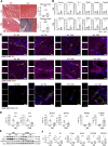
Cardiac fibrosis and innervation. (A) Masson's trichrome staining of young (3‐month‐old) and old (10‐month‐old) WT and mdx mouse hearts. Scale bars, 100 μm. The graph shows the area as a percentage of the fibrosis index (fibrotic area/whole area). N = 3 biological replicates. (B) qPCR for fibrosis‐related transcripts in 3‐ and 10‐month‐old mdx and WT hearts (Col1a1, Col3a1, Fn1, Tgfb1, Twist 1, Twist 2). N = 3 biological replicates. (C) Confocal images for tyrosine hydroxylase (TH), Synapsin 1 (SYN1), MPs (F4/80), proteoglycan CSPG4, and fibronectin 1 (FN1) on myocardium sections of 3‐ and 10‐month‐old mice. The markers TH, SYN1, and CSPG4 are labeled in yellow, cardiac troponin (cTNNT) and F4/80 are in magenta, while FN1 is in white. Scale bars, 10 μm. Nuclei are counterstained with DAPI in blue. (D) Charts indicate positive area expressed as percentage of TH, SYN1, and F4/80 on whole area. N = 3 sections per N = 3 biological replicates. (E and F) Western blot analysis of TH, SYN1, and CSPG4 in mdx and WT hearts of young and old mice. Graphs show optical density (OD) of protein bands normalized to vinculin (VCL). N = 3 biological replicates. Error bars show SEM. *p < 0.05, **p < 0.01, ***p < 0.001, ****p < 0.0001 were calculated using Student's t‐test.
Figure 2
Figure 2
MPs as main producers of CSPG4. (A) Graphic illustration of CSPG4‐CAR‐Ts cells against MPs expressing CSPG4. Created with Biorender.com. Diagrams quantifying the percentage of live cells of bone marrow‐derived MPs of both WT and mdx M0 (nonactivated), M1 (pro‐inflammatory) and M2 (anti‐inflammatory), or CFs after exposure to CSPG4.CARTs compared to cells co‐cultured with control CART (cCART). N = 3 biological replicates. (B) ELISA assay for CSPG4 in supernatants (SUP) isolated from WT and mdx MPs. The chart illustrates the optical density (OD) of CSPG4 in the two groups. Each dot represents the eluted fractions, N = 16. (C) FACSymphony of cardiac tissues. The first chart represents a scatter plot of t‐SNE relative to concatenated total cells from 3‐ or 10‐month‐old WT and mdx hearts. The second chart illustrates the distribution of CD45neg cells (light pink cluster) and myeloid cells (dark pink cluster) in all groups. (D) Graph of FACS analysis relative to number of total MPs (F4/80) positive for CSPG4 in 3‐ or 10‐month‐old WT and mdx hearts. (E) Expression of CSPG4 in subpopulations of MPs: CD80pos (M1), CD206 pos (M2), Ly6Clow (resident), and Ly6Chigh (monocytes) in 3‐ or 10‐month‐old WT and mdx hearts. Error bars show SEM. *p < 0.05, **p < 0.01 calculated using Student's t‐test and one‐way ANOVA.
Figure 3
Figure 3
MPs release CSPG4 in media and inhibit axon growth of dorsal root ganglia. (A) Rendering of 3D NCJ generation. The picture was created with Biorender.com and illustrates the composition of the 3D NCJ model: WT dorsal root ganglia (DRG), WT cardiomyocytes (CMs), and WT CFs were encapsulated in PEG‐FB hydrogel conditioned with the supernatants derived from MP cultures of WT, WT + CSPG4 blocking antibody (BAb), mdx, and mdx + CSPG4 (BAb). (B) Brightfield images of CFs, CMs, and DRG after 1 day of culture (upper panel) and 3D constructs of NCJ models after 7 days (lower panel). Scale bars, 100 μm. (C) Confocal images of 3D NCJ models: WT, WT + CSPG4 BAb, mdx, mdx + CSPG4 BAb. DRG are labeled with tubulin beta 3 (TUBB3, in yellow), CMs with cardiac troponin (cTNNT, in magenta), and CFs with fibroblast marker (ER‐TR7, in white). (D) The graph indicates the area (%) occupied by neuron endings labeled by TUBB3 inside the 3D NCJ models. N = 3 biological replicates, N = 2 sections/constructs. Scale bars, 10 μm. **p < 0.01, ***p < 0.001 were determined using one‐way ANOVA.
Figure 4
Figure 4
Cardiac innervation in old mdx and WT mice. (A) Immunofluorescence staining for tyrosine hydroxylase (TH), Synapsin 1 (SYN1), total MPs (F4/80) and proteoglycan (CSPG4), fibronectin 1 (FN1) on cardiac sections from 10‐month‐old mice. The markers TH and SYN1 are labeled in yellow, while cardiac troponin (cTNNT) and F4/80 are in magenta, FN1 in white, and CSPG4 in yellow or white. DAPI nuclear counterstaining is in blue. Scale bars, 10 μm. (B) Charts indicate area expressed as percentage of TH, SYN1, and F4/80 (positive area/whole area). N = 3 sections per N = 3 biological replicates. (C and D) Western blot analysis for TH, SYN1, and CSPG4 quantified as optical density (OD) of protein bands normalized to vinculin (VCL). N = 3 biological replicates. (E) qPCR relative to markers for neurotransmitter production (Th), survival (Ngf, Ntrk1, and Ngfr) and axon guidance (Tubb3) in DRG. N = 3 biological replicates. Error bars show SEM. *p < 0.05, **p < 0.01, ***p < 0.001, ****p < 0.0001 were calculated using one‐way ANOVA.
Figure 5
Figure 5
Histological analysis and gene profile of cardiac tissue after treatment with givinostat in old mdx and WT mice. (A) Confocal images of immunostaining for gap junctions (CX43) and cardiomyocytes positive for cardiac troponin (cTNNT), N = 3 sections per N = 3 biological replicates. Scale bars, 50 μm. (B) Charts indicate area as percentage of CX43 (positive area/whole area). (C and D) Representative Masson's trichrome images of WT, mdx saline, and mdx Giv hearts and fibrosis index quantification (fibrotic area/whole area) after 60 days of treatment. Scale bars, 100 μm. N = 3 biological replicates. (E) qPCR of fibrosis‐related genes (Tgfb1, Col1a1, Col3a1, Twist1, Twist2, Fn1), cell surface proteoglycan (Cspg4); the enzyme catalyzes the transfer of sulfate groups on CSPG4 chains (Chst11) and inflammatory markers (Adgre1 and Mmp9). N = 3 biological replicates. Error bars show SEM. *p < 0.05, **p < 0.01, ***p < 0.001, ****p < 0.0001 were calculated using one‐way ANOVA and Student's t‐test.
Figure 6
Figure 6
Impact of givinostat on dystrophic cardiomyopathy. (A) Echocardiographic M‐mode images of parasternal long axis view of anterior and posterior walls of left ventricle of WT, mdx saline, and mdx Giv (Vevo 3100, VisualSonics). (B) Echocardiographic parameters: fractional shortening (FS), ejection fraction (EF), and left ventricular end‐diastolic/systolic volume (LV vol;d LV vol;s) in 10‐month‐old mice. The x‐axis shows givinostat or saline treatment on different days (0, 1, 3, 7, 30, and 60). A range of a minimum of three to a maximum of six biological replicates. (C) Hemodynamic analysis including end‐systolic pressure (P es), maximum pressure (P max), minimum rate of pressure change (Min Dp/Dt), and maximum (Max Dp/Dt). N = 3 biological replicates, N = 6 measurements/mouse. Error bars show SEM. *p < 0.05, **p < 0.01, ***p < 0.001, ****p < 0.0001 were calculated using two‐way ANOVA.

Similar articles

Cited by

References

    1. Nowak KJ, Davies KE. Duchenne muscular dystrophy and dystrophin: pathogenesis and opportunities for treatment. EMBO Rep 2004; 5: 872–876. - PMC - PubMed
    1. Lombardi L, Stefano MED, Paggi P. Components of the NGF signaling complex are altered in mdx mouse superior cervical ganglion and its target organs. Neurobiol Dis 2008; 32: 402–411. - PubMed
    1. Shannon TR. Ryanodine receptor Ca2+ sensitivity and excitation‐contraction coupling in muscular dystrophy and heart failure: similar and yet different. Am J Physiol Heart Circ Physiol 2009; 297: H1965–H1966. - PubMed
    1. Canonico F, Chirivi M, Maiullari F, et al. Focus on the road to modelling cardiomyopathy in muscular dystrophy. Cardiovasc Res 2022; 118: 1872–1884. - PubMed
    1. Tripodi L, Villa C, Molinaro D, et al. The immune system in Duchenne muscular dystrophy pathogenesis. Biomedicines 2021; 9: 1447. - PMC - PubMed

Publication types

Substances

LinkOut - more resources