

White Matter Lesion Volumes on 3-T MRI in People With MS Who Had Followed a Diet and Lifestyle Program for More Than 10 Years
- PMID: 39524062
- PMCID: PMC11548950
- DOI: 10.1155/2024/8818934
White Matter Lesion Volumes on 3-T MRI in People With MS Who Had Followed a Diet and Lifestyle Program for More Than 10 Years
Abstract
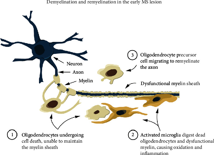
Background: Cerebral white matter lesion (WML) formation in people with multiple sclerosis (pwMS) is linked to the death of myelin-producing oligodendrocytes. Current MS treatment strategies focus on limiting WML accumulation and disability. Using a pathology-supported genetic testing (PSGT) program, we identified specific risk factors for MS, categorized as deficiencies and aggravators. We developed a novel clinical methodology to mitigate these risk factors, including personalized lifestyle interventions and optimization of cerebral nutrients to prevent oligodendrocyte demise and promote remyelination. Objective: To conduct a pilot case-control study over a 10-year period to ascertain whether the PSGT Program can reduce or prevent WML formation in pwMS. Methods: MRI was performed at baseline as well as after an interval period of at least 10 years or longer in 22 pwMS. WML volumes were determined using Sequence Adaptive Multimodal SEGmentation (SAMSEG) software, part of FreeSurfer 7.2. Other variables included age at MRI, disease duration, disability status, and medication. Results: PwMS (n = 13) who had followed the PSGT program for more than 10 years, had significantly smaller lesion volumes (mm3) compared to pwMS who did not adhere to the program (n = 9) (4950 ± 5303 vs. 17934 ± 11139; p = 0.002). WML volumes were significantly associated (p = 0.02) with disability (EDSS) but not with age (p = 0.350), disease duration (p = 0.709), or Interferon-β treatment (p = 0.70). Conclusion: Dietary and lifestyle changes may lower the risk of developing cerebral WMLs in pwMS and potentially slow disease progression. Larger studies are required to confirm the effectiveness of such interventions in pwMS.
Keywords: MRI; SAMSEG; diet; lifestyle; multiple sclerosis; white matter lesion volumes.
Copyright © 2024 Mariaan Jaftha et al.
Conflict of interest statement
Maritha J. Kotze is a nonexecutive director and shareholder of Gknowmix (Pty) Ltd, a spin-out company of the South African Medical Research Council. Lindiwe Whati is a shareholder, and Susan J. van Rensburg is the scientific advisor at Gknowmix (Pty) Ltd. The remaining authors declare no conflicts of interest.
Figures


Similar articles
-
Vascular disease risk factors in multiple sclerosis: Effect on metabolism and brain volumes.Mult Scler Relat Disord. 2023 Jun;74:104675. doi: 10.1016/j.msard.2023.104675. Epub 2023 Mar 31. Mult Scler Relat Disord. 2023. PMID: 37121104 Free PMC article.
-
Association of volumetric MRI measures and disability in MS patients of the same age: Descriptions from a birth year cohort.Mult Scler Relat Disord. 2023 Mar;71:104568. doi: 10.1016/j.msard.2023.104568. Epub 2023 Feb 12. Mult Scler Relat Disord. 2023. PMID: 36805177
-
Pathology-supported genetic testing presents opportunities for improved disability outcomes in multiple sclerosis.Per Med. 2023 Mar;20(2):107-130. doi: 10.2217/pme-2022-0016. Epub 2023 May 17. Per Med. 2023. PMID: 37194915
-
Pathology-supported genetic testing as a method for disability prevention in multiple sclerosis (MS). Part I. Targeting a metabolic model rather than autoimmunity.Metab Brain Dis. 2021 Aug;36(6):1151-1167. doi: 10.1007/s11011-021-00711-w. Epub 2021 Apr 28. Metab Brain Dis. 2021. PMID: 33909200 Review.
-
Pathology-supported genetic testing as a method for disability prevention in multiple sclerosis (MS). Part II. Insights from two MS cases.Metab Brain Dis. 2021 Aug;36(6):1169-1181. doi: 10.1007/s11011-021-00712-9. Epub 2021 Mar 12. Metab Brain Dis. 2021. PMID: 33710528 Review.
References
LinkOut - more resources
Full Text Sources
