

Adipogenesis of bioabsorbable implants under irradiation in a rodent model
- PMID: 39524181
- PMCID: PMC11550574
- DOI: 10.1016/j.reth.2024.10.002
Adipogenesis of bioabsorbable implants under irradiation in a rodent model
Abstract
Background: Breast cancer is the most common cancer among women. Partial mastectomy is an alternative to mastectomy in early-stage breast cancer to restore a poor quality of life (QOL). However, the aesthetic satisfaction with this procedure is inadequate. The standard methods for breast reconstruction have certain limitations. We developed bioabsorbable implants consisting of an outer mesh composed of poly L-lactic acid (PLLA) and an inner component filled with a collagen sponge (CS). These implants were designed to promote and sustain adipogenesis in vivo, without the addition of exogenous cells or growth factors. In this study, we used PLLA mesh implants to investigate the effects of irradiation on fat formation, which is important in partial mastectomy.
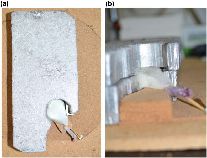
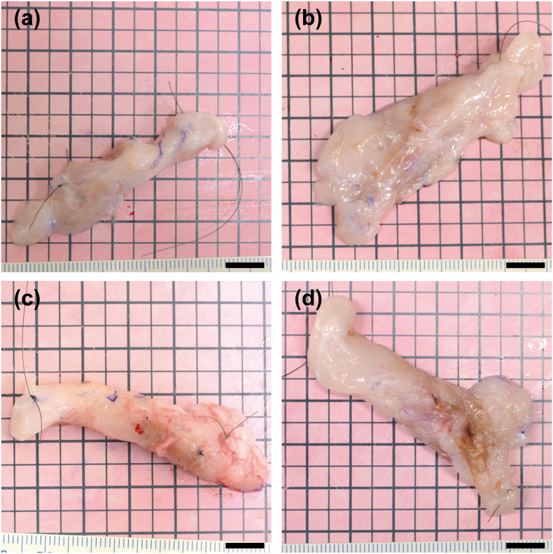
Methods: The implants were inserted into both the inguinal regions of the rats. One month after the implantation, a dose of 13 Gy was delivered to the left-side implants. We compared adipose tissue formation in the non-irradiated and irradiated groups at 6 and 12 months after irradiation.
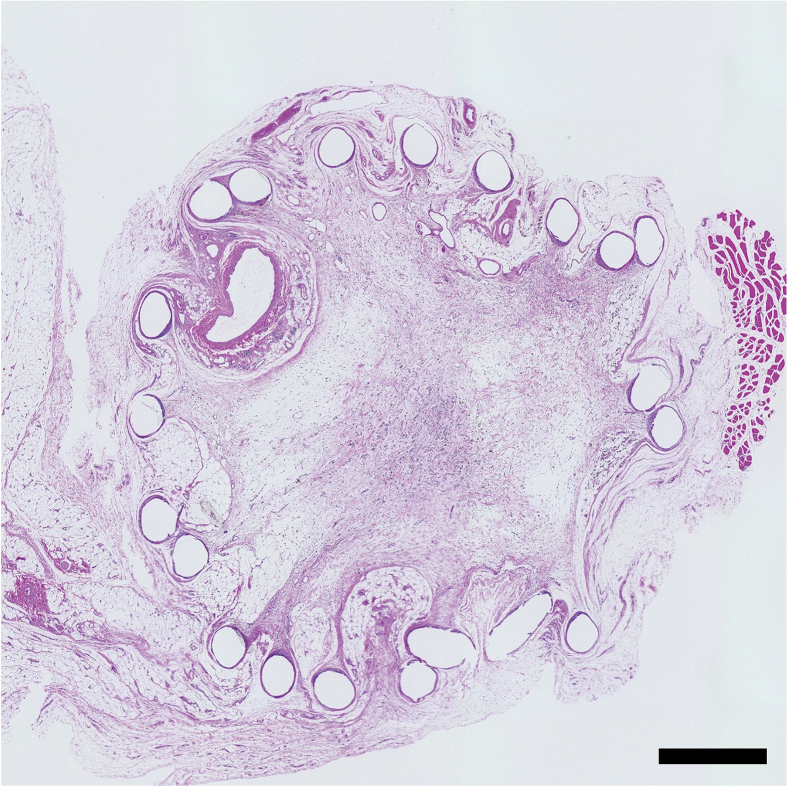
Results: Irradiation of implants did not lead to malignant tumor formation. The newly formed tissues and adipose tissue were not significantly different between the two groups at 6 and 12 months after irradiation.
Conclusions: PLLA mesh implants containing CS are desirable bioabsorbable implants that can be replaced with autologous adipose tissue after in vivo implantation under irradiation. These implants serve as an effective material for partial mastectomy and have the potential to improve the QOL of patients after mastectomy.
Keywords: Adipogenesis; Bioabsorbable; Breast cancer; Irradiation; Partial mastectomy; Poly-L-lactic acid.
© 2024 The Author(s).
Conflict of interest statement
The authors report no proprietary or commercial interest in any product mentioned or concept discussed in this article.
Figures


Similar articles
-
Evaluation of adipogenesis over time using a novel bioabsorbable implant without the addition of exogenous cells or growth factors.Regen Ther. 2024 Jan 4;25:220-228. doi: 10.1016/j.reth.2023.12.015. eCollection 2024 Mar. Regen Ther. 2024. PMID: 38260087 Free PMC article.
-
De novo adipogenesis using a bioabsorbable implant without additional cells or growth factors.J Tissue Eng Regen Med. 2020 Jul;14(7):920-930. doi: 10.1002/term.3041. Epub 2020 Jun 3. J Tissue Eng Regen Med. 2020. PMID: 32293793
-
Development of new bioabsorbable implants with de novo adipogenesis.Regen Ther. 2023 Aug 12;24:311-317. doi: 10.1016/j.reth.2023.07.008. eCollection 2023 Dec. Regen Ther. 2023. PMID: 37638279 Free PMC article.
-
Breast reconstruction with alloplastic implants.J Surg Oncol. 2006 Nov 1;94(6):471-8. doi: 10.1002/jso.20484. J Surg Oncol. 2006. PMID: 17061280 Review.
-
Breast reconstruction after mastectomy.Coll Antropol. 2010 Mar;34 Suppl 1:113-23. Coll Antropol. 2010. PMID: 20402306 Review.
References
-
- Ferlay J., Colombet M., Soerjomataram I., Mathers C., Parkin D.M., Pineros M., et al. Estimating the global cancer incidence and mortality in 2018: GLOBOCAN sources and methods. Int J Cancer. 2019;144:1941–1953. - PubMed
-
- Zainal N.Z., Nik-Jaafar N.R., Baharudin A., Sabki Z.A., Ng C.G. Prevalence of depression in breast cancer survivors: a systematic review of observational studies. Asian Pac J Cancer Prev APJCP. 2013;14:2649–2656. - PubMed
-
- Runowicz C.D., Leach C.R., Henry N.L., Henry K.S., Mackey H.T., Cowens-Alvarado R.L., et al. American cancer society/American society of clinical oncology breast cancer survivorship care guideline. CA Cancer J Clin. 2016;66:43–73. - PubMed
-
- Zhang C., Hu G., Biskup E., Qiu X., Zhang H., Zhang H. Depression induced by total mastectomy, breast conserving surgery and breast reconstruction: a systematic review and meta-analysis. World J Surg. 2018;42:2076–2085. - PubMed
