. 2024 Oct 11;27(11):111163.
doi: 10.1016/j.isci.2024.111163. eCollection 2024 Nov 15.

Tau processing and tau-mediated inflammation differ in human APOEε2 and APOEε4 astrocytes

Affiliations

Tau processing and tau-mediated inflammation differ in human APOEε2 and APOEε4 astrocytes

Tobias Mothes et al. iScience. .

Abstract

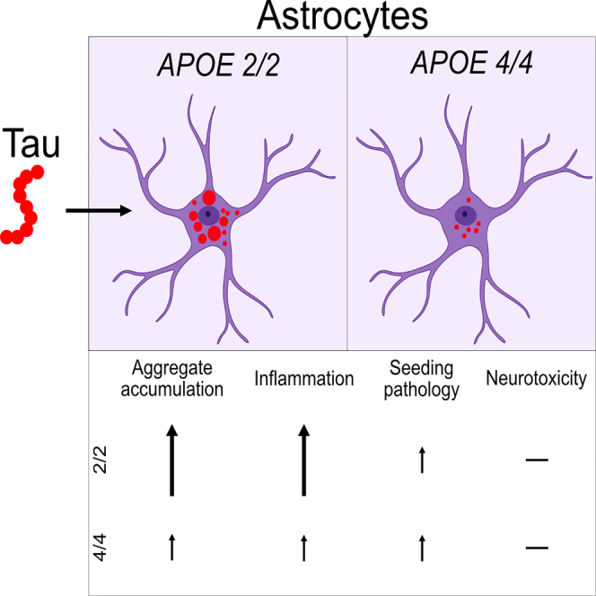
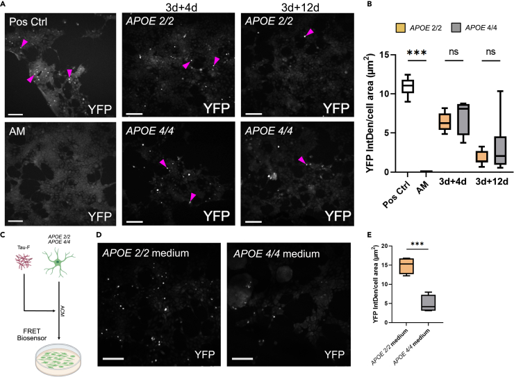
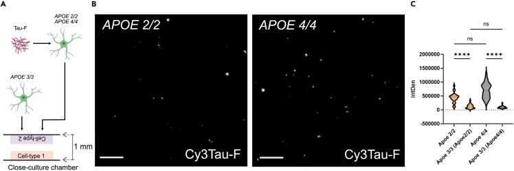
Alzheimer's disease (AD) and progressive supra-nuclear palsy (PSP) are both proteinopathies, characterized by the accumulation of tau aggregates. APOEε4 is the greatest genetic risk factor for developing AD, while APOEε2 is a significant risk factor for developing PSP. In the brain, astrocytes are the predominant producer of ApoE, but they are also important for inflammation and overall brain homeostasis. Although, tau inclusions appear frequently in astrocytes in both AD and PSP brains, their connection to ApoE remains unclear. Here, we show that hiPSC-derived APOE 2/2 astrocytes accumulate, process, and spread pathogenic tau aggregates more efficiently than isogenic APOE 4/4 astrocytes. Moreover, the APOE 2/2 astrocytes display a more robust inflammatory response, which could be of relevance for the disease course. Taken together, our data highlight a central role of ApoE in astrocyte-mediated tau pathology.

Keywords: disease; genes; neuroscience.


Conflict of interest statement

The authors declare that no competing interests.

Figures

None
Graphical abstract
Figure 1
Figure 1
Characterization of APOE 2/2 and APOE 4/4 astrocytes and the experimental setup (A) DNA analysis confirming successful gene editing of BIONi308-A-2 (APOE 2/2) and BIONi037-A-(APOE 4/4) cells. (B) Example images of control APOE 2/2 and APOE 4/4 astrocytes, stained for vimentin and GFAP. Scale bars = 100 μm. (C) Schematic outline of the experimental setup. Isogenic human iPSC derived astrocytes (APOE 2/2 and APOE 4/4) were exposed to synthetic tau fibrils (Tau-F) for 3 days. Then, the astrocytes were washed and further incubated in a tau-free medium for 4–12 days. Cells were fixed or lysed at 3days+4days and 3days + 12days and medium was collected at 4, 8, and 12 days post-wash. The cells were analyzed using immunocytochemistry, Western blot and cultured in a close-culture setup together with APOE 3/3 astrocytes. The medium was directly analyzed using IP, ELISA and a cytokine array (MSD-ECL); or added to iPSC derived neuronal cultures or HEK293T RD tau FRET Biosensor c (seeding assay). (D) z stack of Cy3Tau-F inclusions inside APOE 2/2 and APOE 4/4 astrocyte at 3days+4days, vimentin (white), tau (red) and DAPI (blue). Scale bar = 50 μm.
Figure 2
Figure 2
APOE genotype drastically affects processing of internalized tau protein in iPSC derived astrocytes (A) Representative images of Cy3 labeled Tau-F internalized by APOE 2/2 and APOE 4/4 astrocytes at 3 + 4 and 3 + 12 days (The same images, including membrane dye and DAPI are shown in Figure S3). Scale bars = 100 μm. (B) Quantification of intracellular Cy3Tau-F IntDen, normalized to number of nuclei per field. Analyzed using one-way ANOVA. (C–E) Number of small, medium, and large Cy3Tau-F inclusions inside APOE 2/2 and APOE 4/4 astrocytes at 3days+4days and 3days + 12days. Small aggregates = 0.53–21.1 μm2, medium aggregates = 21.2–105.6 μm2 and large aggregates = 105.7–1056 μm2. (F) Total tau (Tau-12, Tau-5, BT2, and T46) Western blot analysis of the soluble fraction of lysed APOE 2/2 and APOE 4/4 astrocytes at 3 + 12days. (G) Quantification of the WB in f. Band intensity was normalized to the band intensity of the NoStain blot (Figure S5). Analyzed using a standard t-test. (H) Total tau (Tau-12, Tau-5, BT2, and T46) western blot of the insoluble fraction from APOE 2/2 and APOE 4/4 astrocytes lysates after 3 + 12days exposure. Band 1–75 kDa; Band 2–60 kDa. (I) Quantification of WB in h. Band intensity was normalized to the band intensity of the NoStain blot (Figure S5). Analyzed using a standard t-test. (J) Sandwich ELISA analysis of ACM from APOE 2/2 and APOE 4/4 astrocytes at the different time points (3days+4days, 3days+8days and 3days + 12days). (K) Sandwich ELISA analysis of lysates from APOE 2/2 and APOE 4/4 astrocytes. (L) Tau concentration measured by ELISA in the ApoE-IP fraction of ACM from Tau-F exposed APOE 2/2 and APOE 4/4 astrocytes. (M) WB of ApoE-IP fraction stained using the same ApoE antibody (NoStain blot in Figure S5). Data is presented as mean ± SD. P-values are presented as following; ∗p < 0.05, ∗∗p < 0.01, ∗∗∗p < 0.005, ∗∗∗∗p < 0.0001.
Figure 3
Figure 3
APOE genotype influences the inflammatory response of iPSC derived astrocytes (A) Quantification of vimentin expression in APOE 2/2 and APOE 4/4 astrocytes, normalized to the number of nuclei per field. (B) Quantification of GFAP expression in APOE 2/2 and APOE 4/4 astrocytes, normalized to the number of nuclei per field. (C–H) Cytokine levels in ACM from control/Tau-F exposed APOE 2/2 and APOE 4/4 astrocytes as measured by U-Plex MesoScale. (C) IL-8. (D) IL-12/IL-23p40. (E) CXCL11. (F) CCL2. (G) CXCL10. (H) TNF-α. Two-way ANOVA with multiple comparisons between all groups was used for statistical analysis for each cytokine. Data are presented as mean ± SD. P-values are presented within each time point; ∗p < 0.05, ∗∗p < 0.01, ∗∗∗p < 0.005, ∗∗∗∗p < 0.0001.
Figure 4
Figure 4
Seeding competent Tau-F aggregates are excreted from astrocytes of both genotypes but seeding is more efficient in the presence of ApoE 2 RD tau P301S FRET biosensor cells treated with ACM from Tau-F exposed APOE 2/2 or APOE 4/4 astrocytes. Positive control is the starting medium (same as the astrocytes were exposed to at day 0), and negative control is the astrocyte medium without tau (AM). All medium was fortified with 1% lipofectamine and 10% FBS. (A) Representative YFP images of Biosensor cells after 48h exposure to ACM from the first and last time point (3days+4days ACM, 3days + 12days ACM) plus positive and negative controls. The purple arrows indicate examples of positive puncta. Scale bars = 100 μm. (B) Quantification of YFP IntDen normalized to the cell area. Analyzed using one-way ANOVA with multiple comparisons between APOE 2/2 and APOE 4/4 for each time point. (C) Schematic figure of the experimental layout. Tau-F was diluted in ACM from control astrocytes and added to FRET biosensor cells. (D) Representative YFP images of Biosensor cells exposed to tau diluted in ACM from APOE 2/2 (APOE 2/2 medium) and APOE 4/4 astrocytes (APOE 4/4 medium). Scale bars = 100 μm. (E) Quantification of YFP IntDen, normalized to the cell area. Data are presented as mean ± SD. P-values are presented as following; ∗p < 0.05, ∗∗p < 0.01, ∗∗∗p < 0.005, ∗∗∗∗p < 0.0001.
Figure 5
Figure 5
Close-culture experiments, indicate that presence of ApoE 3 influences the Tau processing in APOE 2/2 and APOE 4/4 astrocytes (A) Schematic outline of the experimental layout. APOE 2/2 and APOE 4/4 astrocytes were exposed to Tau-F before being placed in a “close-culture” chamber with APOE 3/3 astrocytes. (B) Representative images of intracellular Cy3Tau-F signal in APOE 2/2 and APOE 4/4 astrocytes after four days of close-culturing (the complete images for all groups and an overview in gray-scale are shown in Figures S8 and S9). Scale bars = 100 µm. (C) Quantification of tau deposits in the astrocytes (APOE 2/2 and APOE 4/4 with their corresponding APOE 3/3 astrocytes), intracellular Cy3Tau-F IntDen, normalized to the number of nuclei per field. Data are analyzed using one-way ANOVA with multiple comparisons between each group. Data are presented as mean ± SD. P-values are presented as following; ∗p < 0.05, ∗∗p < 0.01, ∗∗∗p < 0.005, ∗∗∗∗p < 0.0001.
Figure 6
Figure 6
ACM from APOE 2/2 and APOE 4/4 astrocytes does not have an immediate effect on neuronal health (A) Schematic outline of the experimental layout. Neurons were cultured for two weeks with ACM from either APOE 2/2 or APOE 4/4 astrocytes (with or without tau inclusions). Addition of a regular astrocyte medium (not conditioned) was used as a control. Neuronal health was assessed using an LDH assay at d7 and d14, as well as an ATP assay at d14. (B) Example images of control neurons at day 28 of differentiation (d0 of exposure), stained for βIII-tubulin, MAP2, synaptophysin, and tau. Scale bars = 100μm. Quantification of LDH assay for (C) neurons and (D) astrocytes. Assay was performed on the medium from neurons and astrocytes at 7days and 14days (prior to medium change). Values are plotted as LDH enzymatic activity (nmol/min/mL). (E) Quantification of ATP in neurons after 14 days of exposure to ACM from APOE 2/2 or APOE 4/4 astrocytes, with or without tau. Regular astrocyte medium was included as a control (Ctrl). Two-way ANOVA with multiple comparisons for all mean values was used for statistical analysis. Data is presented as mean ± SD. P-values are presented as following; ∗p < 0.05, ∗∗p < 0.01, ∗∗∗p < 0.005, ∗∗∗∗p < 0.0001.

Similar articles

Cited by

References

    1. Yoshida M. Astrocytic inclusions in progressive supranuclear palsy and corticobasal degeneration. Neuropathology. 2014;34:555–570. doi: 10.1111/neup.12143. - DOI - PubMed
    1. Komori T., Arai N., Oda M., Nakayama H., Mori H., Yagishita S., Takahashi T., Amano N., Murayama S., Murakami S., et al. Astrocytic plaques and tufts of abnormal fibers do not coexist in corticobasal degeneration and progressive supranuclear palsy. Acta Neuropathol. 1998;96:401–408. doi: 10.1007/s004010050911. - DOI - PubMed
    1. Richetin K., Steullet P., Pachoud M., Perbet R., Parietti E., Maheswaran M., Eddarkaoui S., Bégard S., Pythoud C., Rey M., et al. Tau accumulation in astrocytes of the dentate gyrus induces neuronal dysfunction and memory deficits in Alzheimer’s disease. Nat. Neurosci. 2020;23:1567–1579. doi: 10.1038/s41593-020-00728-x. - DOI - PubMed
    1. Sofroniew M.V., Vinters H.V. Astrocytes: biology and pathology. Acta Neuropathol. 2010;119:7–35. doi: 10.1007/s00401-009-0619-8. - DOI - PMC - PubMed
    1. Liao F., Yoon H., Kim J. Apolipoprotein E metabolism and functions in brain and its role in Alzheimer’s disease. Curr. Opin. Lipidol. 2017;28:60–67. doi: 10.1097/MOL.0000000000000383. - DOI - PMC - PubMed

LinkOut - more resources