. 2024 Oct 25:15:1472656.
doi: 10.3389/fimmu.2024.1472656. eCollection 2024.

Human genital dendritic cell heterogeneity confers differential rapid response to HIV-1 exposure

Affiliations

Human genital dendritic cell heterogeneity confers differential rapid response to HIV-1 exposure

Siddharth Parthasarathy et al. Front Immunol. .

Abstract

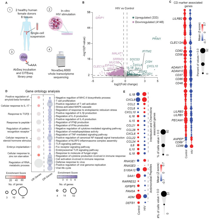
Dendritic cells (DCs) play critical roles in HIV pathogenesis and require further investigation in the female genital tract, a main portal of entry for HIV infection. Here we characterized genital DC populations at the single cell level and how DC subsets respond to HIV immediately following exposure. We found that the genital CD11c+HLA-DR+ myeloid population contains three DC subsets (CD1c+ DC2s, CD14+ monocyte-derived DCs and CD14+CD1c+ DC3s) and two monocyte/macrophage populations with distinct functional and phenotypic properties during homeostasis. Following HIV exposure, the antiviral response was dominated by DCs' rapid secretory response, activation of non-classical inflammatory pathways and host restriction factors. Further, we uncovered subset-specific differences in anti-HIV responses. CD14+ DCs were the main population activated by HIV and mediated the secretory antimicrobial response, while CD1c+ DC2s activated inflammasome pathways and IFN responses. Identification of subset-specific responses to HIV immediately after exposure could aid targeted strategies to prevent HIV infection.

Keywords: HIV; antiviral response; dendritic cell; female genital tract; mucosal immunity; single-cell RNA sequencing.


Conflict of interest statement

The authors declare that the research was conducted in the absence of any commercial or financial relationships that could be construed as a potential conflict of interest.

Figures

Figure 1
Figure 1
Identification of phenotypically and transcriptionally unique DC subsets in the genital tract. (A) Representative gating strategy to identify FGT resident CD11c+HLA-DR+ cells using a combination of surface protein expression (AbSeq) and RNA expression in CITEseq dataset. Y-axis and x-axis indicate normalized AbSeq expression levels. (B) Representative UMAP visualization of FGT resident CD11c+HLA-DR+ cells through unbiased clustering, depicting expression of discriminating RNA expression (left column) and surface protein expression (right column) in distinct subsets, Cluster 1 (blue), Cluster 2 (red), Cluster 3 (teal) and Cluster 4 (green). (C) Scatter plot comparing surface antibody expression between different FGT CD11c+HLA-DR+ subsets. (D) AUCell analysis of top 50 genes in DC3 cluster described by AC Villani et. al.
Figure 2
Figure 2
Functional specialization of genital DCs under homeostatic conditions. (A) Hierarchical clustering heatmap comparing gene expression of significant pattern recognition receptors (PRRs) between FGT resident CD11c+HLA-DR+ subsets. (B) Hierarchical clustering heatmap comparing gene expression of top 5 interleukin and chemokines in each cluster, where size of the circle indicates percentage of cells expressing associated gene > 0 (C) AUCell comparison of key gene ontology (GO) terms associated with dendritic cell function between DC subsets.
Figure 3
Figure 3
CD14 DCs represent a heterogenous, activated population within the genital mucosa. (A) Representative flow cytometry gating strategy to identify FGT resident CD11c+HLA-DR+ cells. (B) Representative plot of HLA-DR+CD11c+ subsets based on CD14 and CD1c expression; CD14+CD1c- (I, orange), CD14-CD1c+ (II, red), CD14+CD1c+ (III, green) and CD14-CD1c- (IV, pink). (C) Comparison of CD11c+HLA-DR+ subset frequencies in the FGT. (D) Comparison of CD11c+HLA-DR+ subset distribution across different anatomical regions of the FGT, endometrium (EM), endocervix (END) and ectocervix (ECX). (E) Representative gating to differentiate CD14-CD1c- cells based on CD16 expression (left); Histogram comparing CLEC12A expression between CD14-CD16+ and CD14-CD16- cells (right). (F) Comparison of surface protein expression between different CD11c+HLA-DR+ cells; HLA-DRhigh (top left), CD64 (top middle), CD11b (top right), CD54 (bottom left), CLEC12A (bottom middle) and CX3CR1 (bottom left). (G) Comparison of HIV tropic surface proteins between different CD11c+HLA-DR+ cells; CD4 (top left), CXCR4 (top right), CCR5 (bottom right) and CD49d (bottom right). Statistics – Friedman’s multiple comparison, (p ≤ 0.05 *; p ≤ 0.01 **; p ≤ 0.001 ***; p ≤ 0.0001 ****).
Figure 4
Figure 4
Genital DCs undergo rapid transcriptional changes in response to HIV stimulation. (A) Graphical depiction of CITEseq protocol to identify transcriptional changes induced by HIV in FGT resident CD11c+HLA-DR+ cells. Mixed single-cell suspensions from hysterectomy samples were incubated with HIV at MOI of 0.5 for 30 minutes at 37°C (B) Volcano plot of significantly upregulated (n=333) and downregulated genes (n=4148) by HIV in CD11c+HLA-DR+ cells. (C) Hierarchical clustering heatmap comparing gene expression of CD markers between control and HIV exposed cells. (D) Bubble-plot visualization of significant GO terms enriched in genes upregulated (UR genes; red) and downregulated (DR genes; blue) in response to HIV by CD11c+HLA-DR+ cells. (E) Hierarchical clustering heatmap comparing genes expression of secreted factors such as interleukins, chemokines and antimicrobial proteins between control and HIV exposed CD11c+HLA-DR+ cells. Statistics – Volcano plot, non-parametric ANOVA (p ≤ 0.05; -1.2 <FC>1.2); GO terms significance of FDR ≤ 0.05.
Figure 5
Figure 5
CD14+ DCs largely mediate rapid protective host response observed in genital DC response to HIV. (A) Representative UMAP visualization of CD11c+HLA-DR+ clusters and (B) overlay of control and HIV exposed cells (MOI=0.5 for 30 min). (C) AUCell comparing enrichment in CD11c+HLA-DR+ subsets in HIV exposed samples of overall upregulated (UR) and downregulated (DR) HIV signatures (top row) and key gene ontology (GO) terms associated with dendritic cell function (bottom row). (D) Venn-diagram representation comparing expression of shared and unique genes significantly upregulated (top) and downregulated (bottom) between CD11c+HLA-DR+ subsets in response to HIV stimulation. (p-value ≤ 0.05; -1.2 <FC>1.2) (E) Bubble-plot representation of biological pathways significantly Reactome significantly enriched in upregulated (UR; red) and downregulated (DR; blue) genes in response to HIV stimulation between different CD11c+HLA-DR+ subsets. (FDR ≤ 0.05; black dots indicate non-significant (n.s.) FDR values) (F) Comparison of secreted protein levels in supernatants of media (control; blue) and HIV exposed (HIV; red; MOI 0.5 for 3h), FGT resident CD14+ cells. Significance – Paired non-parametric Wilcoxon-test (p ≤ 0.05 *; p ≤ 0.01 **).

References

    1. Leung Soo C, Pant Pai N, Bartlett SJ, Esmail A, Dheda K, Bhatnagar S. Socioeconomic factors impact the risk of HIV acquisition in the township population of South Africa: A Bayesian analysis. PloS Glob Public Health. (2023) 3:e0001502. doi: 10.1371/journal.pgph.0001502 - DOI - PMC - PubMed
    1. Collaborators GH. Global, regional, and national sex-specific burden and control of the HIV epidemic 1990-2019, for 204 countries and territories: the Global Burden of Diseases Study 2019. Lancet HIV. (2021) 8:e633–51. doi: 10.1016/S2352-3018(21)00152-1 - DOI - PMC - PubMed
    1. How is HIV transmitted (2021). Available online at: https://www.nichd.nih.gov/health/topics/hiv/conditioninfo/transmission (Accessed May 29 2024).
    1. Nazli A, Chan O, Dobson-Belaire WN, Ouellet M, Tremblay MJ, Gray-Owen SD, et al. . Exposure to HIV-1 directly impairs mucosal epithelial barrier integrity allowing microbial translocation. PloS Pathog. (2010) 6:e1000852. doi: 10.1371/journal.ppat.1000852 - DOI - PMC - PubMed
    1. Ferreira VH, Dizzell S, Nazli A, Kafka JK, Mueller K, Nguyen PV, et al. . Medroxyprogesterone acetate regulates HIV-1 uptake and transcytosis but not replication in primary genital epithelial cells, resulting in enhanced T-cell infection. J Infect Dis. (2015) 211:1745–56. doi: 10.1093/infdis/jiu832 - DOI - PubMed