. 2024 Nov 6:19:11397-11413.
doi: 10.2147/IJN.S470465. eCollection 2024.

Temperature and Ultrasound-Responsive Nanoassemblies for Enhanced Organ Targeting and Reduced Cardiac Toxicity

Affiliations

Temperature and Ultrasound-Responsive Nanoassemblies for Enhanced Organ Targeting and Reduced Cardiac Toxicity

Mingzhou Jiang et al. Int J Nanomedicine. .

Abstract

Background: Biocompatible nanocarriers are widely employed as drug-delivery vehicles for treatment. Nevertheless, indiscriminate drug release, insufficient organ-specific targeting, and systemic toxicity hamper nanocarrier effectiveness. Stimuli-responsive nano-sized drug delivery systems (DDS) are an important strategy for enhancing drug delivery efficiency and reducing unexpected drug release.

Methods: This study introduces a temperature- and ultrasound-responsive nano-DDS in which the copolymer p-(MEO2MA-co-THPMA) is grafted onto mesoporous iron oxide nanoparticles (MIONs) to construct an MPL-p nano-DDS. The copolymer acts as a nanopore gatekeeper, assuming an open conformation at sub-physiological temperatures that allows drug encapsulation and a closed conformation at physiological temperatures that prevents unexpected drug release during circulation. Lactoferrin was conjugated to the nanoparticle surface via polyethylene glycol to gain organ-targeting ability. External ultrasonic irradiation of the nanoparticles in the targeted organs caused a conformational change of the copolymer and reopened the pores, facilitating controlled drug release.

Results: MPL-p exhibited excellent biocompatibility and rare drug release in circulation. When targeting delivery to the brain, ultrasound promoted the release of the loaded drugs in the brain without accumulation in other organs, avoiding the related adverse reactions, specifically those affecting the heart.

Conclusion: This study established a novel temperature- and ultrasound-responsive DDS that reduced systemic adverse reactions compared with traditional DDS, especially in the heart, and demonstrated excellent organ delivery efficiency.

Keywords: drug delivery; mesoporous iron oxide nanoparticles; systemic toxicity; temperature- and ultrasound-responsive.


Conflict of interest statement

The authors report no conflicts of interest in this work.

Figures

Figure 1
Figure 1
Synthesis and working schematic diagram of the novel brain-targeted nano ultrasound-controlled release system DOX-MPL-p. At temperatures below LCST, the drug is loaded into MPL-p. At temperatures higher than LCST, DOX-MPL-P blocks the drug and is injected intravenously into mice, limiting the release of DOX in the circulation. With LF, DOX-MPL-p targets the brain across BBB in a receptor-mediated mechanism and eventually disrupts to release of the drug upon ultrasonic stimulation.
Figure 2
Figure 2
Procedures of synthesizing the MION-PEG-LF-p(MEO2MA-co-THPMA). MIONs went through sequential conjugation with LF, Mal-PEG-NHS, and p(MEO2MA-co-THPMA) to get MION-PEG-LF- p(MEO2MA-co-THPMA), which was named as MPL-p.
Figure 3
Figure 3
Characterization of MION-PEG-LF-p(MEO2MA-co-THPMA) during synthesis and drug loading process. The TEM images of MION (a); (b) Pore size distribution of MION; (c) N2 adsorption/desorption isotherm of MION; The TEM images of DOX-MPL-p (d); (e) The DLS results for DOX-MION; (f) The DLS results for DOX-MPL-p; (g) Stability of DOX-MION and DOX-MPL-p in physiological saline, n=3; (h) Comparison of average weight of MION, MION-PEG-LF and MPL-p, n=6; (i) Fluorescence spectra of DOX and DOX-MPL-p with a DOX concentration of 0.5 mg/mL. Asterisks were used to illustrate the level of significance: (***) for P < 0.001.
Figure 4
Figure 4
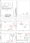
Characterization of MION-PEG-LF-p(MEO2MA-co-THPMA). (a) 1H NMR spectra of p(MEO2MA-co-THPMA) before and after ultrasound treated for 10min, 1 MHz, 100W; (b) Compare the infrared spectra of MION and MION-PEG-LF in the range of 400–4000 cm−1. The vibration of C-O in PEG alcohol molecules was observed at 1100 cm−1; (c) Compare the infrared spectra of MION and MION-PEG-LF in the range of 400–800 cm−1. The typical C-S bond in MION-PEG-LF was observed at 669 cm−1, which is evidence of the conjugation of thiolated LF; (d) The infrared spectrum of the copolymer p(MEO2MA-co-THPMA) shows a characteristic peak of THPMA at 1104 cm−1 and a characteristic peak of MEO2MA at 1723 cm−1; (e) Compare the infrared spectra of MION-PEG-LF and MION-PEG-LF-p(MEO2MA-co-THPMA) in the range of 400–4000 cm−1. The vibration of C-O in PEG alcohol molecules and the characteristic peak of THPMA were observed at 1100 cm−1, the characteristic peak of MEO2MA was observed at 1722 cm−1, and the vinyl functional group on MION was observed at 1650 cm−1, which is the key group for connecting the copolymer to MION.
Figure 5
Figure 5
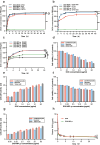
Toxicity assessment and drug release characterization of DOX MPL in vivo and in vitro. Drug release rates of DOX-MPL-p and DOX-MION in vitro at different temperatures (a) and ultrasound-stimulated environment (b); (c) Drug release rates of DOX-MPL-p at different ultrasonic frequencies; Cell viability (d) and lactate dehydrogenase (LDH) activity (e) of cardiomyocytes, neurons and tumor cell after administration of DOX; Cell viability (f) and lactate dehydrogenase (LDH) activity (g) of cardiomyocytes, neurons and tumor cell after administration of DOX-MPL-p; (h) Temporal variations in plasma DOX concentrations of free DOX and DOX-MPL-p post-injection. Asterisks were used to illustrate the level of significance: (**) for P < 0.01, (***) for P < 0.001, and (ns) for P > 0.05.
Figure 6
Figure 6
Biosafety of DOX-MPL-p. (a) The H&E staining results of the vital organs of mice injected with DOX-MPL-p observed at 40x magnification, revealed no significant pathological damage. This observation was consistent across various time points: 24 h, 7 d, 14 d, and 21 d post-administration; (b) Heart rates and artery pressures (c) in 16 h after injection. Asterisks were used to illustrate the level of significance: (*) for P < 0.05, (**) for P < 0.01.
Figure 7
Figure 7
MPL-p reduces the damage of drugs to cardiac function as a DDS. (a) Survival estimates by Kaplan–Meier in Vehicle (PBS), DOX, and DOX-MPL-p treatment groups within 30 d (n = 8); (b) Representation images of H&E staining in mice heart after drug treatment; (c) Quantification of heart weight/tibia length; (d) Representative M-mode echocardiographic images of each group at 14 d point after drug treatment; (e) stroke volume, ejection fraction (f), and fraction shortening (g) measured by echocardiography. Asterisks were used to illustrate the level of significance: (*) for P < 0.05, (**) for P < 0.01, and (***) for P < 0.001.
Figure 8
Figure 8
Organ-specific drug release properties of DOX-MPL-p. (a) Fluorescence signals of the brain, heart, liver, spleen, and kidney presented at 0.5 h, 4 h, 8 h, and 24 h after injection of DiR@MPL-p and DiR@MION; (b) Immunofluorescence images of the frozen brain tissues at 24 h after injection of DiR@MPL-p and DiR@MION, and the white arrows indicates the red fluorescent signal emitted by DiR@MPL-p in the brain tissue; Compare the intensity of fluorescence signal in liver (c) and brain (d); (e) The DOX content in the brain tissue by ultrasonic stimulation at 2 h, 4 h, and 8 h after administration of DOX-MPL-p. Asterisks were used to illustrate the level of significance: (*) for P < 0.05, (**) for P < 0.01, and (***) for P < 0.001.

Similar articles

Cited by

References

    1. Jensen SA, Day ES, Ko CH, et al. Spherical nucleic acid nanoparticle conjugates as an RNAi-based therapy for glioblastoma. Sci Transl Med. 2013;5(209):209ra152. doi:10.1126/scitranslmed.3006839 - DOI - PMC - PubMed
    1. Greish K. Enhanced permeability and retention (EPR) effect for anticancer nanomedicine drug targeting. Methods Mol Biol. 2010;624:25–37. - PubMed
    1. Maeda H, Wu J, Sawa T, Matsumura Y, Hori K. Tumor vascular permeability and the EPR effect in macromolecular therapeutics: a review. J Control Release. 2000;65(1–2):271–284. doi:10.1016/S0168-3659(99)00248-5 - DOI - PubMed
    1. Christidi E, Brunham LR. Regulated cell death pathways in doxorubicin-induced cardiotoxicity. Cell Death Dis. 2021;12(4):339. doi:10.1038/s41419-021-03614-x - DOI - PMC - PubMed
    1. Mura S, Nicolas J, Couvreur P. Stimuli-responsive nanocarriers for drug delivery. Nat Mater. 2013;12(11):991–1003. doi:10.1038/nmat3776 - DOI - PubMed

LinkOut - more resources