. 2024 Nov 4:19:11257-11273.
doi: 10.2147/IJN.S481044. eCollection 2024.

Exosomes Derived from Antler Mesenchymal Stem Cells Promote Wound Healing by miR-21-5p/STAT3 Axis

Affiliations

Exosomes Derived from Antler Mesenchymal Stem Cells Promote Wound Healing by miR-21-5p/STAT3 Axis

Deshuang Meng et al. Int J Nanomedicine. .

Abstract

Background: Deer antlers, unique among mammalian organs for their ability to regenerate annually without scar formation, provide an innovative model for regenerative medicine. This study explored the potential of exosomes derived from antler mesenchymal stem cells (AMSC-Exo) to enhance skin wound healing.

Methods: We explored the proliferation, migration and angiogenesis effects of AMSC-Exo on HaCaT cells and HUVEC cells. To investigate the skin repairing effect of AMSC-Exo, we established a full-thickness skin injury mouse model. Then the skin thickness, the epidermis, collagen fibers, CD31 and collagen expressions were tested by H&E staining, Masson's trichrome staining and immunofluorescence experiments. MiRNA omics analysis was conducted to explore the mechanism of AMSC-Exo in skin repairing.

Results: AMSC-Exo stimulated the proliferation and migration of HaCaT cells, accelerated the migration and angiogenesis of HUVEC cells. In the mouse skin injury model, AMSC-Exo stimulated angiogenesis and regulated the extracellular matrix by facilitating the conversion of collagen type III to collagen type I, restoring epidermal thickness to normal state without aberrant hyperplasia. Notably, AMSC-Exo enhanced the quality of wound healing with increased vascularization and reduced scar formation. MiRNAs in AMSC-Exo, especially through the miR-21-5p/STAT3 signaling pathway, played a crucial role in these processes.

Conclusion: This study underscores the efficacy of AMSC-Exo in treating skin wounds, suggesting a new approach for enhancing skin repair and regeneration.

Keywords: deer antler; exosomes; mesenchymal stem cells; microRNA; skin wound healing.


Conflict of interest statement

The authors report no conflicts of interest in this work.

Figures

Figure 1
Figure 1
Isolation and identification of the AMSC. (A) The tip of velvet antler, black arrow indicates the reserve mesenchymal layer. (B) Optical morphology of AMSC under light field microscope, scale bars: 500 μm. The expressions of AMSC surface markers CD44 and CD90 identified by immunofluorescence staining (C) and flow cytometry (D), scale bar: 100 μm. Adipogenic, osteogenic and chondrogenic differentiation potentials of AMSC examined by oil red O (E), scale bar: 100 μm, alizarin red (F), scale bar: 200 μm and alcian blue staining (G), scale bar: 500 μm.
Figure 2
Figure 2
The characterization of AMSC-Exo. (A) TEM image of AMSC-Exo, scale bar: 200 nm. (B) Immunoblot analysis of known exosomal markers (CD81, TSG101 and ALIX) and negative marker (GM130) in AMSC-Exo. The particle size distribution of AMSC-Exo detected by nanoflow cytometry (C) and nanocoulter counter (D). (E) The zeta potential of AMSC-Exo measured by nanocoulter counter.
Figure 3
Figure 3
Effects of AMSC-Exo on cell proliferation, migration and tubule formation. (A) The cell viability of HaCaT cells after 72 h treatment with AMSC-Exo. (B) HaCaT cell migration was detected by scratch assay, scale bar: 500 μm. (C) Cell migration area was counted. (D) HUVEC cell migration was detected by transwell, scale bar: 100 μm. (E) HUVEC transmembrane cell number was counted. (F) The tube formation of HUVEC, scale bar: 500 μm. HUVEC tubule formation node (G), junction number (H) and tube length (I). In the picture *Means p < 0.05, **Means p < 0.005, and ***Means p < 0.001.
Figure 4
Figure 4
The effect of AMSC-Exo on wound healing in a mouse full-thickness skin injury model. (A) Experimental procedure of the mice. (B) Global morphological photographs of wound area treated with PBS (Ctrl), gel (Gel) or gel-AMSC-Exo (G-Exo) on day 0, 7, 16, 22, 29. scale bar: 5 mm. (C) Wound area ratios of day 29 to day 0. In the picture *Means p < 0.05 and ***Means p < 0.001.
Figure 5
Figure 5
AMSC-Exo improved the healing quality of back wound in mice. (A) Representative images of H&E, Masson’s trichrome staining and CD31 immunohistochemistry of the skin sections of day 29 in Ctrl group, Gel group or G-Exo group. The red triangles indicate the CD31 positive expression of new blood vessels, scale bar: 50 μm. (B) Quantitative analysis of the thickness of newly formed epidermis. (C) Number of CD31 positive stained vessels. (D) COL1 and COL3 immunohistochemical staining of the sections on day 29 after different treatments, scale bar: 50 μm. (E) Quantitative analysis of COL3 and COL1. In the picture *Means p < 0.05, **Means p < 0.005, and ****Means p < 0.0001.
Figure 6
Figure 6
AMSC-Exo miRNA sequencing and target gene cluster analysis. (A) Top 20 miRNAs expressed in AMSC-Exo. (B) GO enrichment barplot. (C) Top 20 of biological process enrichment. (D) Top 20 of KEGG enrichment.
Figure 7
Figure 7
STAT3 is the direct target of miR-21-5p. (A) Prediction of miR-21-5p target genes by TargetScan. (B) The binding sites between miR-21-5p and STAT3. (C) Dual luciferase reporter gene analysis in HaCaT cells. (D) Relative protein expression of STAT3, TIMP3, MMP1 and GAPDH in HaCaT cells after miR-21-5p mimics (miR-21) or miR-21-5p inhibitor (miR-21i) treatment. In the picture **Means p < 0.005, and ***Means p < 0.001, ****Means p < 0.0001.
Figure 8
Figure 8
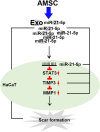
Schematic diagram of the experimental results.

Similar articles

Cited by

References

    1. Zouboulis CC, Makrantonaki E. Clinical aspects and molecular diagnostics of skin aging. Clin Dermatol. 2011;29(1):3–14. doi:10.1016/j.clindermatol.2010.07.001 - DOI - PubMed
    1. Farage MA, Miller KW, Elsner P, et al. Characteristics of the aging skin. Adv Wound Care. 2013;2(1):5–10. doi:10.1089/wound.2011.0356 - DOI - PMC - PubMed
    1. Li Q, Gong S, Yao W, et al. Exosome loaded genipin crosslinked hydrogel facilitates full thickness cutaneous wound healing in rat animal model. Drug Delivery. 2021;28(1):1. - PMC - PubMed
    1. Cha J, Falanga V. Stem cells in cutaneous wound healing. Clin Dermatol. 2007;25(1):73–78. doi:10.1016/j.clindermatol.2006.10.002 - DOI - PubMed
    1. Burd A, Ahmed K, Lam S, et al. Stem cell strategies in burns care. Burns. 2007;33(3):282–291. doi:10.1016/j.burns.2006.08.031 - DOI - PubMed

LinkOut - more resources